Cell Biochemistry and Biophysics ( IF 1.8 ) Pub Date : 2022-10-06 , DOI: 10.1007/s12013-022-01105-0 Bhawantha M Jayawardena 1 , Resmi Menon 1, 2 , Mark R Jones 1 , Christopher E Jones 1
|
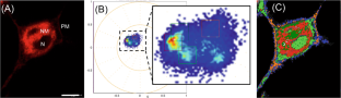
The interaction of protein and peptide amyloid oligomers with membranes is thought to be one of the mechanisms contributing to cellular toxicity. However, techniques to study these interactions in the complex membrane environment of live cells are lacking. Spectral phasor analysis is a recently developed biophysical technique that can enable visualisation and analysis of membrane-associated fluorescent dyes. When the spectral profile of these dyes changes as a result of changes to the membrane microenvironment, spectral phasor analysis can localise those changes to discrete membrane regions. In this study, we investigated whether spectral phasor analysis could detect changes in the membrane microenvironment of live cells in the presence of fibrillar aggregates of the disease-related Aβ42 peptide or the functional amyloid neurokinin B. Our results show that the fibrils cause distinct changes to the microenvironment of nile red associated with both the plasma and the nuclear membrane. We attribute these shifts in nile red spectral properties to changes in membrane fluidity. Results from this work suggest that cells have mechanisms to avoid or control membrane interactions arising from functional amyloids which have implications for how these peptides are stored in dense core vesicles. Furthermore, the work highlights the utility of spectral phasor analysis to monitor microenvironment changes to fluorescent probes in live cells.
中文翻译:
尼罗红的光谱相量分析确定了淀粉样肽存在下的膜微环境变化
蛋白质和肽淀粉样蛋白寡聚体与膜的相互作用被认为是导致细胞毒性的机制之一。然而,缺乏研究活细胞复杂膜环境中这些相互作用的技术。光谱相量分析是最近开发的生物物理技术,可以实现膜相关荧光染料的可视化和分析。当这些染料的光谱曲线因膜微环境的变化而发生变化时,光谱相量分析可以将这些变化定位到离散的膜区域。在这项研究中,我们研究了光谱相量分析是否可以检测在存在疾病相关 Aβ 42纤维状聚集体的情况下活细胞膜微环境的变化肽或功能性淀粉样神经激肽 B。我们的结果表明,原纤维会导致与血浆和核膜相关的尼罗红微环境发生明显变化。我们将尼罗红光谱特性的这些变化归因于膜流动性的变化。这项工作的结果表明,细胞具有避免或控制由功能性淀粉样蛋白引起的膜相互作用的机制,这对这些肽如何储存在致密核心囊泡中具有影响。此外,该工作强调了光谱相量分析在监测活细胞中荧光探针的微环境变化方面的实用性。


 京公网安备 11010802027423号