Skeletal Radiology ( IF 1.9 ) Pub Date : 2022-09-28 , DOI: 10.1007/s00256-022-04190-7 Brian Lue 1 , Joshua Amaya 1 , Flavio Duarte Silva 1 , Katherine Raspovic 2 , Yin Xi 1 , Avneesh Chhabra 1, 2, 3, 4, 5
|
Background
Foot and ankle amputation is a feared complication of diabetic neuropathy and diabetes mellitus (DM) accounts for 80% of all in-hospital amputations. Magnetic resonance neurography is an effective tool in characterizing neuromuscular sequelae of the disease. However, conventional ankle MRI is more commonly performed and has not been studied to assess neuromuscular changes of DM.
Objective
The objective is to characterize neuromuscular changes of diabetic patients in a case–control study using conventional ankle MRI.
Methods
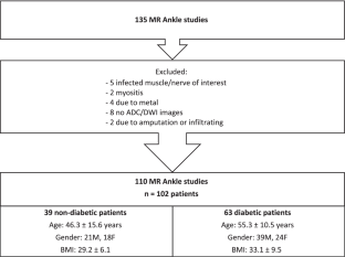
Between November 2019 and July 2021, 110 consecutive ankle MRI scans (n = 102 patients) at our county hospital were reviewed and met the inclusion criteria. Patients were divided into two cohorts, diabetic (N = 63) and non-diabetic (N = 39). Demographics, HgbA1c, and reason for MRI study were collected via retrospective chart review. The presence of intramuscular edema-like signal, pattern of the edema, muscle fatty infiltration, and measurements of the cross-sectional area of the posterior, medial, and lateral tibial nerves (PTN, MPN, and LPN) was recorded blinded to the clinical findings by two readers.
Results
Muscle edema-like signal was much more likely to be found in DM (odds ratio 19.5, 95% CI 7.0–54.6, p < 0.001). DM also showed increase of 0.87 in the mean grade of muscle fatty infiltration (p < 0.001). There were higher rates of nerve T2 hyperintensity (odds ratio 14.0, 95% CI 3.1–62.7, p < 0.001) and the measured areas of the PTN, MPN, and LPN were also larger in DM compared to their non-diabetic counterparts (PTN: 0.16 cm2 vs. 0.10 cm2, p < 0.01; MPN: 0.09 cm2 vs. 0.05 cm2, p < 0.01; LPN: 0.07 cm2 vs. 0.04 cm2, p < 0.05).
Conclusion
Conventional ankle MRIs can be used to detect DM-related neuromuscular changes.
中文翻译:
表征糖尿病患者神经和肌肉变化的常规踝关节 MRI 结果:病例对照研究
背景
足踝截肢是糖尿病性神经病变的一种可怕并发症,糖尿病 (DM) 占所有住院截肢手术的 80%。磁共振神经造影是表征该疾病的神经肌肉后遗症的有效工具。然而,传统的踝关节 MRI 更常被执行,并且尚未被研究用于评估 DM 的神经肌肉变化。
客观的
目的是在使用传统踝关节 MRI 的病例对照研究中表征糖尿病患者的神经肌肉变化。
方法
2019 年 11 月至 2021 年 7 月期间,对我们县医院进行的 110 例连续踝关节 MRI 扫描(n = 102 例患者)进行了审查并符合纳入标准。患者被分为两组,糖尿病患者(N = 63)和非糖尿病患者(N = 39)。通过回顾性图表审查收集人口统计学、HgbA1c 和进行 MRI 研究的原因。记录肌内水肿样信号的存在、水肿模式、肌肉脂肪浸润,以及胫后神经、内侧神经和外侧神经(PTN、MPN 和 LPN)横截面积的测量结果,这些测量结果对临床试验不知情两位读者的发现。
结果
肌肉水肿样信号更可能出现在 DM 中(比值比 19.5,95% CI 7.0–54.6,p < 0.001)。DM 还显示肌肉脂肪浸润的平均等级增加了 0.87 ( p < 0.001)。神经 T2 高信号率更高(比值比 14.0,95% CI 3.1–62.7,p < 0.001),与非糖尿病患者相比,DM 的 PTN、MPN 和 LPN 的测量面积也更大(PTN :0.16 cm 2对比 0.10 cm 2,p < 0.01;MPN:0.09 cm 2对比 0.05 cm 2,p < 0.01;LPN:0.07 cm 2对比 0.04 cm 2,p < 0.05)。
结论
传统的踝关节 MRI 可用于检测 DM 相关的神经肌肉变化。


 京公网安备 11010802027423号