Skeletal Radiology ( IF 1.9 ) Pub Date : 2022-09-20 , DOI: 10.1007/s00256-022-04183-6 Chandana Kurra 1 , Paul Wasserman 1 , Anthony Khoury 2 , Michael Freidl 3
|
Objective
To offer an adjunctive imaging tool to MRI for evaluating tape suture related rotator cuff repairs.
Materials and methods
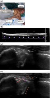
A two-part pilot study was performed to assess visibility of tape suture following imaging with various modalities. Institutional research ethics board approval was obtained prior to cadaveric studies. Two tape sutures, FiberTape® and TigerTape®, were evaluated in each experiment. The first experiment assessed the tape suture’s presence in a gelatin mold following exposure to X-ray, ultrasound, computed tomography (CT), and magnetic resonance imaging (MRI) without contrast. The second experiment assessed tape suture’s visibility in a cadaveric shoulder model following a standard of care, double-row, transosseous equivalent rotator cuff repair. The same imaging protocols and equipment were used for each part of the study with the addition of MR arthrography–tailored images on the cadaveric specimen. All images were assessed by a musculoskeletal trained radiologist.
Results
The gelatin study demonstrated that the tape suture was visible via ultrasound only. X ray, CT, and MRI did not show tape suture material. In the ultrasound component of the cadaveric study, distinct echogenic textural detail of the tape suture was easily identified, compatible with the simulated rotator cuff repair. X ray and unenhanced CT did not show the tape suture material or the rotator cuff. MRI without intraarticular gadolinium contrast did not adequately image the suture tape; however, faint artifact in the repair region was visualized. MRI with intra-articular contrast did not show the tape suture material in detail; however, the intraarticular gadolinium did provide an advantageous background of high T1 signal that contrasted with the cuff/suture construct.
Conclusion
Ultrasound proved to be an effective imaging modality to visualize tape suture in both the gelatin and cadaveric parts of the pilot study. Ultrasound may be a useful tool to evaluate post-operative tape suture–related repairs in patients that cannot obtain MRIs or when the MRI findings are equivocal.
中文翻译:
胶带缝线的成像特征:一种现代手术材料
客观的
为 MRI 提供辅助成像工具,用于评估胶带缝合相关的肩袖修复。
材料和方法
进行了一项分为两部分的试点研究,以评估用各种模式成像后胶带缝合的可见性。在尸体研究之前获得了机构研究伦理委员会的批准。在每个实验中评估了两种胶带缝合线,FiberTape® 和 TigerTape®。第一个实验评估了在暴露于 X 射线、超声波、计算机断层扫描 (CT) 和无对比磁共振成像 (MRI) 后胶带缝合线在明胶模具中的存在。第二个实验评估了根据护理标准、双排、经骨等效肩袖修复,胶带缝合在尸体肩部模型中的可见性。研究的每个部分都使用了相同的成像协议和设备,并在尸体标本上添加了 MR 关节造影定制图像。
结果
明胶研究表明,胶带缝合线仅通过超声波可见。X 射线、CT 和 MRI 未显示胶带缝合材料。在尸体研究的超声成分中,很容易识别胶带缝合的明显回声纹理细节,与模拟肩袖修复兼容。X 射线和未增强 CT 未显示胶带缝合材料或肩袖。没有关节内对比剂的 MRI 不能充分显示缝合带;但是,可以看到修复区域中的微弱伪影。关节内造影 MRI 未详细显示胶带缝合材料;然而,关节内钆确实提供了高 T1 信号的有利背景,这与袖带/缝线构造形成对比。
结论
超声被证明是一种有效的成像方式,可以在试验研究的明胶和尸体部分可视化胶带缝合。对于无法获得 MRI 图像或 MRI 结果不明确的患者,超声可能是评估术后胶带缝合相关修复的有用工具。


 京公网安备 11010802027423号