Nature Biomedical Engineering ( IF 26.8 ) Pub Date : 2022-09-19 , DOI: 10.1038/s41551-022-00940-z Rui Cao 1 , Scott D Nelson 2 , Samuel Davis 1 , Yu Liang 3 , Yilin Luo 1 , Yide Zhang 1 , Brooke Crawford 4 , Lihong V Wang 1
|
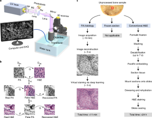
Obtaining frozen sections of bone tissue for intraoperative examination is challenging. To identify the bony edge of resection, orthopaedic oncologists therefore rely on pre-operative X-ray computed tomography or magnetic resonance imaging. However, these techniques do not allow for accurate diagnosis or for intraoperative confirmation of the tumour margins, and in bony sarcomas, they can lead to bone margins up to 10-fold wider (1,000-fold volumetrically) than necessary. Here, we show that real-time three-dimensional contour-scanning of tissue via ultraviolet photoacoustic microscopy in reflection mode can be used to intraoperatively evaluate undecalcified and decalcified thick bone specimens, without the need for tissue sectioning. We validate the technique with gold-standard haematoxylin-and-eosin histology images acquired via a traditional optical microscope, and also show that an unsupervised generative adversarial network can virtually stain the ultraviolet-photoacoustic-microscopy images, allowing pathologists to readily identify cancerous features. Label-free and slide-free histology via ultraviolet photoacoustic microscopy may allow for rapid diagnoses of bone-tissue pathologies and aid the intraoperative determination of tumour margins.
中文翻译:
通过深度学习辅助紫外光声显微镜对骨组织进行无标记术中组织学研究
获得骨组织冰冻切片用于术中检查具有挑战性。因此,为了确定切除的骨边缘,骨科肿瘤学家依靠术前 X 射线计算机断层扫描或磁共振成像。然而,这些技术无法进行准确诊断或术中确认肿瘤边缘,并且在骨肉瘤中,它们可能导致骨边缘比必要的宽 10 倍(体积上 1,000 倍)。在这里,我们表明,通过紫外光声显微镜在反射模式下对组织进行实时三维轮廓扫描可用于术中评估未脱钙和脱钙厚骨标本,而无需进行组织切片。我们通过传统光学显微镜获得的金标准苏木精和伊红组织学图像验证了该技术,并且还表明,无监督的生成对抗网络实际上可以对紫外光声显微镜图像进行染色,使病理学家能够轻松识别癌症特征。通过紫外光声显微镜进行的无标记和无载玻片组织学可以快速诊断骨组织病理,并有助于术中确定肿瘤边缘。


 京公网安备 11010802027423号