Neuroradiology ( IF 2.4 ) Pub Date : 2022-09-19 , DOI: 10.1007/s00234-022-03052-2 Zhaoting Meng 1, 2 , Lingyu Zhang 1 , Caiyun Huang 3 , Yingshi Piao 4, 5 , Xiaohong Chen 6 , Junfang Xian 1
|
Purpose
Among head and neck cancers, hypopharyngeal squamous cell carcinoma (HSCC) shows the highest malignancy, which is associated with histologic grading. This study was designed to investigate whether quantitative parameters derived from 18F-fluorodeoxyglucose positron emission tomography/magnetic resonance imaging (18F-FDG PET/MRI) can preoperatively estimate the histologic grade of HSCC.
Methods
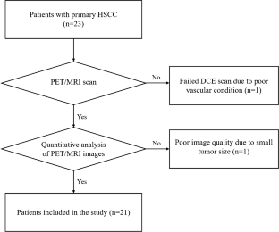
18F-FDG PET/MRI of neck was successfully performed in 21 patients with histologically proven HSCC including poorly differentiated group (ten patients) and well-moderately differentiated group (eleven patients). Quantitative parameters derived from FDG-PET, diffusion-weighted imaging (DWI), and dynamic contrast enhanced-magnetic resonance imaging (DCE-MRI) were calculated based on volume of interest drawn on the tumor and compared between two groups. The efficacy of quantitative parameters for the estimation of histologic grades of HSCC was evaluated.
Results
There were statistically significant differences in mean value of standard uptake value (SUV), apparent diffusion coefficient (ADC), and Ktrans derived from 18F-FDG PET/MRI of HSCC between two groups (p < 0.05). There was no statistically significant difference in other quantitative parameters derived from 18F-FDG PET/MRI of HSCC between two groups. The area under the curve (AUC) of the combination of SUVmean, ADCmean, and Ktrans in the estimation of histologic grade of HSCC was 0.936 with sensitivity of 90.0% and specificity of 81.8%.
Conclusion
The combination of SUVmean, ADCmean, and Ktrans derived from 18F-FDG PET/MRI can accurately predict the histologic grade of HSCC preoperatively.
中文翻译:
18F-氟脱氧葡萄糖正电子发射断层扫描/磁共振成像定量参数可准确估计术前下咽鳞状细胞癌的组织学分级
目的
在头颈癌中,下咽鳞状细胞癌 (HSCC) 的恶性程度最高,这与组织学分级有关。本研究旨在探讨来自18 F-氟脱氧葡萄糖正电子发射断层扫描/磁共振成像 ( 18 F-FDG PET/MRI) 的定量参数是否可以在术前估计 HSCC 的组织学分级。
方法
21例经组织学证实为HSCC的患者成功进行颈部18 F-FDG PET/MRI检查,包括低分化组(10例)和中高分化组(11例)。FDG-PET、弥散加权成像 (DWI) 和动态对比增强磁共振成像 (DCE-MRI) 的定量参数基于在肿瘤上绘制的感兴趣体积计算并在两组之间进行比较。评估了定量参数估计 HSCC 组织学分级的功效。
结果
两组HSCC 18 F-FDG PET/MRI标准摄取值(SUV)、表观扩散系数(ADC)、 K trans平均值比较差异有统计学意义( p < 0.05)。两组HSCC 18 F-FDG PET/MRI的其他定量参数差异无统计学意义。SUV均值、ADC均值和K trans联合估计HSCC组织学分级的曲线下面积(AUC)为0.936,敏感性为90.0%,特异性为81.8%。
结论
18 F-FDG PET/MRI得出的SUV mean、ADC mean和K trans的组合可以准确预测HSCC术前的组织学分级。


 京公网安备 11010802027423号