Acta Neuropathologica ( IF 9.3 ) Pub Date : 2022-08-30 , DOI: 10.1007/s00401-022-02477-6 Abris Gilvesy 1, 2 , Evelina Husen 1 , Zsofia Magloczky 3 , Orsolya Mihaly 4 , Tibor Hortobágyi 5, 6, 7, 8 , Shigeaki Kanatani 9 , Helmut Heinsen 10, 11 , Nicolas Renier 12 , Tomas Hökfelt 1 , Jan Mulder 1 , Mathias Uhlen 1, 13 , Gabor G Kovacs 14, 15 , Csaba Adori 1
|
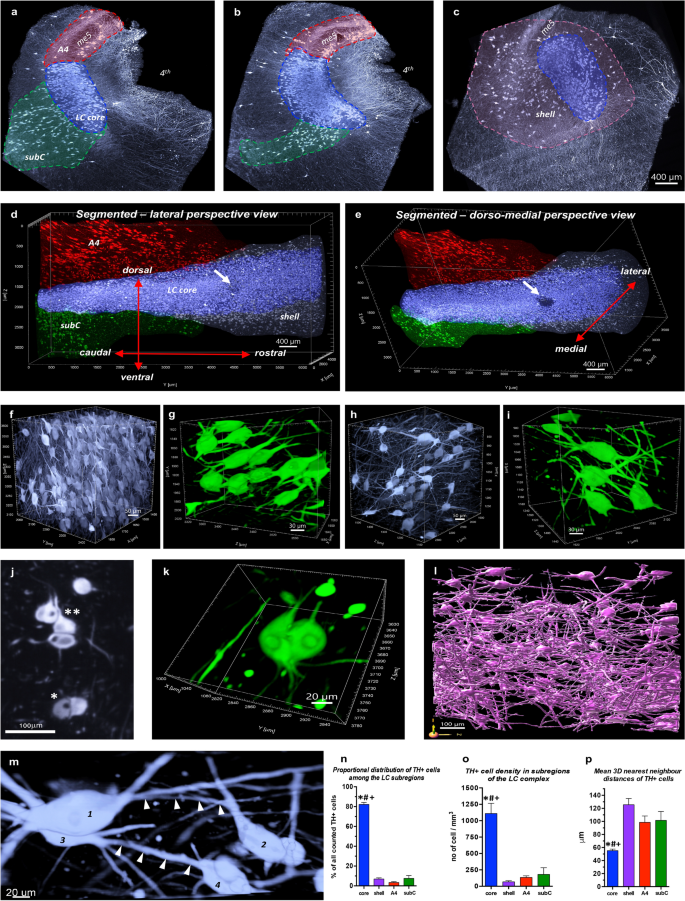
Tau pathology of the noradrenergic locus coeruleus (LC) is a hallmark of several age-related neurodegenerative disorders, including Alzheimer’s disease. However, a comprehensive neuropathological examination of the LC is difficult due to its small size and rod-like shape. To investigate the LC cytoarchitecture and tau cytoskeletal pathology in relation to possible propagation patterns of disease-associated tau in an unprecedented large-scale three-dimensional view, we utilized volume immunostaining and optical clearing technology combined with light sheet fluorescence microscopy. We examined AT8+ pathological tau in the LC/pericoerulear region of 20 brains from Braak neurofibrillary tangle (NFT) stage 0–6. We demonstrate an intriguing morphological complexity and heterogeneity of AT8+ cellular structures in the LC, representing various intracellular stages of NFT maturation and their diverse transition forms. We describe novel morphologies of neuronal tau pathology such as AT8+ cells with fine filamentous somatic protrusions or with disintegrating soma. We show that gradual dendritic atrophy is the first morphological sign of the degeneration of tangle-bearing neurons, even preceding axonal lesions. Interestingly, irrespective of the Braak NFT stage, tau pathology is more advanced in the dorsal LC that preferentially projects to vulnerable forebrain regions in Alzheimer’s disease, like the hippocampus or neocortical areas, compared to the ventral LC projecting to the cerebellum and medulla. Moreover, already in the precortical Braak 0 stage, 3D analysis reveals clustering tendency and dendro-dendritic close appositions of AT8+ LC neurons, AT8+ long axons of NFT-bearing cells that join the ascending dorsal noradrenergic bundle after leaving the LC, as well as AT8+ processes of NFT-bearing LC neurons that target the 4th ventricle wall. Our study suggests that the unique cytoarchitecture, comprised of a densely packed and dendritically extensively interconnected neuronal network with long projections, makes the human LC to be an ideal anatomical template for early accumulation and trans-neuronal spreading of hyperphosphorylated tau.
中文翻译:
通过三维成像对人类蓝斑-蓝周复合体中细胞 tau 病理学的时空表征
去甲肾上腺素能蓝斑 (LC) 的 Tau 病理学是多种与年龄相关的神经退行性疾病(包括阿尔茨海默病)的标志。然而,由于 LC 体积小且呈杆状,因此很难对其进行全面的神经病理学检查。为了在前所未有的大规模三维视图中研究与疾病相关 tau 的可能传播模式相关的 LC 细胞结构和 tau 细胞骨架病理学,我们利用体积免疫染色和光学清除技术结合光片荧光显微镜。我们检查了来自 Braak 神经原纤维缠结 (NFT) 阶段 0-6 的 20 个大脑的 LC/pericoerulear 区域中的 AT8 +病理性 tau。我们展示了 AT8 +有趣的形态复杂性和异质性LC 中的细胞结构,代表 NFT 成熟的各个细胞内阶段及其不同的过渡形式。我们描述了神经元 tau 病理学的新形态,例如 AT8 +具有细丝状体细胞突起或具有崩解体细胞的细胞。我们表明逐渐的树突萎缩是缠结神经元退化的第一个形态学标志,甚至在轴突损伤之前。有趣的是,无论 Braak NFT 阶段如何,与投射到小脑和髓质的腹侧 LC 相比,tau 病理学在背侧 LC 中更先进,优先投射到阿尔茨海默病的脆弱前脑区域,如海马或新皮质区域。此外,已经在皮质前 Braak 0 阶段,3D 分析揭示了 AT8 + LC 神经元、AT8 +离开 LC 后加入上行背侧去甲肾上腺素能束的携带 NFT 的细胞的长轴突,以及靶向第四脑室壁的携带 NFT 的 LC 神经元的AT8 +突。我们的研究表明,独特的细胞结构,由密集堆积和树突状广泛互连的神经元网络组成,具有长投影,使人类 LC 成为过度磷酸化 tau 的早期积累和跨神经元扩散的理想解剖模板。


 京公网安备 11010802027423号