Acta Pharmacologica Sinica ( IF 6.9 ) Pub Date : 2022-08-25 , DOI: 10.1038/s41401-022-00967-7 Sun-Li Hu 1, 2, 3 , Abdullah Al Mamun 2 , Jian Shaw 1, 2, 3 , Sun-Long Li 3 , Yi-Feng Shi 3 , Xue-Man Jin 3 , Ying-Xin Yu 3 , Chao-Zhi Pang 3 , Ze-Yang Li 3 , Jia-Jie Lu 3 , Yue-Piao Cai 1, 2 , Xiang-Yang Wang 1, 2, 3 , Jian Xiao 1, 2
|
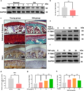
Mitochondrial dynamics, including mitochondrial fission and fusion, are critical for maintaining mitochondrial functions. Evidence shows that TANK-binding kinase 1 (TBK1) regulates mitochondrial fusion and fission and then mitophagy. Since a previous study demonstrates a strong correlation between mitophagy and osteoarthritis (OA), we herein investigated the potential role of TBK1 in OA process and mitochondrial functions. We demonstrated a strong correlation between TBK1 and OA, evidenced by significantly downregulated expression of TBK1 in cartilage tissue samples of OA patients and in the chondrocytes of aged mice, as well as TNF-α-stimulated phosphorylation of TBK1 in primary mouse chondrocytes. TBK1 overexpression significantly attenuated TNF-α-induced apoptosis and abnormal mitochondrial function in primary mouse chondrocytes. Furthermore, TBK1 overexpression induced remodeling of mitochondrial morphology by directly phosphorylating dynamin-related protein 1 (DRP1) at Ser637, abolishing the fission of DRP1 and preventing its fragmentation function. Moreover, TBK1 recruitment and DRP1 phosphorylation at Ser637 was necessary for engulfing damaged mitochondria by autophagosomal membranes during mitophagy. Moreover, we demonstrated that APMK/ULK1 signaling contributed to TBK1 activation. In OA mouse models established by surgical destabilization of the medial meniscus, intraarticular injection of lentivirus-TBK1 significantly ameliorated cartilage degradation via regulation of autophagy and alleviation of cell apoptosis. In conclusion, our results suggest that the TBK1/DRP1 pathway is involved in OA and pharmacological targeting of the TBK1-DRP1 cascade provides prospective therapeutic benefits for the treatment of OA.
中文翻译:
TBK1 介导的 DRP1 磷酸化协调骨关节炎中的线粒体动力学和自噬激活
线粒体动力学,包括线粒体裂变和融合,对于维持线粒体功能至关重要。有证据表明 TANK 结合激酶 1 (TBK1) 调节线粒体融合和裂变,然后调节线粒体自噬。由于先前的研究表明线粒体自噬与骨关节炎 (OA) 之间存在很强的相关性,因此我们在此研究了 TBK1 在 OA 过程和线粒体功能中的潜在作用。我们证明了 TBK1 和 OA 之间存在很强的相关性,OA 患者软骨组织样本和老年小鼠软骨细胞中 TBK1 表达显着下调,以及原代小鼠软骨细胞中 TNF-α 刺激的 TBK1 磷酸化证明了这一点。 TBK1 过表达显着减弱原代小鼠软骨细胞中 TNF-α 诱导的细胞凋亡和线粒体功能异常。此外,TBK1过表达通过直接磷酸化动力相关蛋白1(DRP1)Ser637位点诱导线粒体形态重塑,消除DRP1的裂变并阻止其断裂功能。此外,TBK1 募集和 DRP1 Ser637 磷酸化对于线粒体自噬过程中自噬体膜吞噬受损线粒体是必要的。此外,我们证明 APMK/ULK1 信号传导有助于 TBK1 激活。在通过手术破坏内侧半月板建立的 OA 小鼠模型中,关节内注射慢病毒-TBK1 通过调节自噬和减轻细胞凋亡,显着改善软骨退化。总之,我们的结果表明 TBK1/DRP1 通路参与 OA,并且 TBK1-DRP1 级联的药理学靶向为 OA 的治疗提供了前瞻性的治疗益处。


 京公网安备 11010802027423号