当前位置:
X-MOL 学术
›
Microsc. Res. Tech.
›
论文详情
Our official English website, www.x-mol.net, welcomes your
feedback! (Note: you will need to create a separate account there.)
Developmental events in the lung of the Japanese quail (Coturnix coturnix japonica): Morphological, histochemical and electron-microscopic studies
Microscopy Research and Technique ( IF 2.0 ) Pub Date : 2022-08-24 , DOI: 10.1002/jemt.24225 Heba Mostafa 1 , Manal T Hussein 2 , Mahmoud Abd-Elnaeim 1
Microscopy Research and Technique ( IF 2.0 ) Pub Date : 2022-08-24 , DOI: 10.1002/jemt.24225 Heba Mostafa 1 , Manal T Hussein 2 , Mahmoud Abd-Elnaeim 1
Affiliation
|
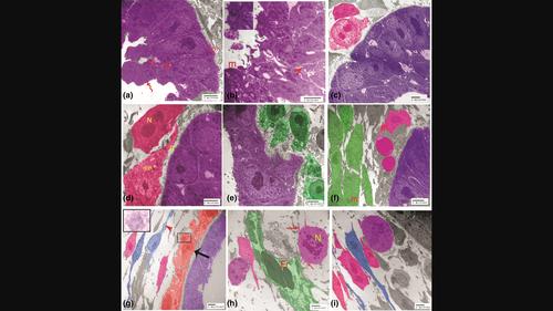
The lung of birds is the most complex and efficient gas exchanger in the air-breathing vertebrates. A total number of 27 normal Japanese quail embryos during the pre-hatching period were used. The current work aimed to investigate the histomorphological, histochemical and ultrastructural changes of the lung at different stages of development using light and electron microscopy. The results showed that the respiratory primordium was observed on the 2nd embryonic day (ED) as a ventral out-pouching of the primitive foregut into the surrounding mesenchyme. The first evidence of the lung buds appeared on the 3rd ED. On the 4th ED, the buds increased in size. In addition, the secondary bronchi budded from the epithelial lining of the primary bronchus into the surrounding mesenchyme. On the 6th ED, the number of the secondary bronchi increased and began to give rise small tubules (parabronchi). On the 7th ED, telocytes (TCs) were seen extensively in relation to the undifferentiated mesenchymal cells (UMC) and surrounding the developing bronchi and blood vessels. On the 8th ED, the number of the small-diameter, smooth-walled parabronchi (PR) was considerably increased. On the 9th ED, anastomosing of the developing parabronchi was observed forming large diameter ones and the open-type endocrine cells in the parabronchial wall were observed. On the 10th ED, deep costal impressions on the lung tissue were seen, the atrial primordia could be observed and the primordia of all air sac were developed. In conclusion; morphogenesis and differentiation of the developing lung epithelium depend on epithelial-mesenchyme interaction.
中文翻译:

日本鹌鹑 (Coturnix coturnix japonica) 肺的发育事件:形态学、组织化学和电子显微镜研究
鸟类的肺是呼吸空气的脊椎动物中最复杂和最有效的气体交换器。在孵化前阶段,总共使用了 27 个正常的日本鹌鹑胚胎。目前的工作旨在使用光学和电子显微镜研究肺在不同发育阶段的组织形态学、组织化学和超微结构变化。结果表明,在胚胎第 2 天 (ED) 观察到呼吸原基是原始前肠向周围间充质的腹侧外翻。肺芽的第一个证据出现在第 3 个 ED。在第 4 个 ED,芽的大小增加了。此外,次级支气管从初级支气管的上皮衬里出芽进入周围的间充质。在 6 日,次级支气管的数量增加并开始产生小管(副支气管)。在第 7 个 ED,在未分化的间充质细胞 (UMC) 和发育中的支气管和血管周围广泛地看到了 telocytes (TCs)。在第 8 个 ED,小直径、壁光滑的副支气管 (PR) 的数量显着增加。第9天,观察到发育中的支气管旁支气管吻合形成大直径支气管旁支气管,支气管旁壁内可见开放型内分泌细胞。10日,可见肺组织深肋压痕,可见心房原基,所有气囊原基均已发育。综上所述; 发育中的肺上皮的形态发生和分化取决于上皮-间充质相互作用。
更新日期:2022-08-24
中文翻译:
日本鹌鹑 (Coturnix coturnix japonica) 肺的发育事件:形态学、组织化学和电子显微镜研究
鸟类的肺是呼吸空气的脊椎动物中最复杂和最有效的气体交换器。在孵化前阶段,总共使用了 27 个正常的日本鹌鹑胚胎。目前的工作旨在使用光学和电子显微镜研究肺在不同发育阶段的组织形态学、组织化学和超微结构变化。结果表明,在胚胎第 2 天 (ED) 观察到呼吸原基是原始前肠向周围间充质的腹侧外翻。肺芽的第一个证据出现在第 3 个 ED。在第 4 个 ED,芽的大小增加了。此外,次级支气管从初级支气管的上皮衬里出芽进入周围的间充质。在 6 日,次级支气管的数量增加并开始产生小管(副支气管)。在第 7 个 ED,在未分化的间充质细胞 (UMC) 和发育中的支气管和血管周围广泛地看到了 telocytes (TCs)。在第 8 个 ED,小直径、壁光滑的副支气管 (PR) 的数量显着增加。第9天,观察到发育中的支气管旁支气管吻合形成大直径支气管旁支气管,支气管旁壁内可见开放型内分泌细胞。10日,可见肺组织深肋压痕,可见心房原基,所有气囊原基均已发育。综上所述; 发育中的肺上皮的形态发生和分化取决于上皮-间充质相互作用。


 京公网安备 11010802027423号