The Ocular Surface ( IF 5.9 ) Pub Date : 2022-08-20 , DOI: 10.1016/j.jtos.2022.07.008 Anam Akhlaq 1 , Clara Colón 2 , Bernardo M Cavalcanti 2 , Shruti Aggarwal 2 , Yureeda Qazi 2 , Andrea Cruzat 3 , Candice Jersey 2 , Douglas B Critser 4 , Amy Watts 2 , Jill Beyer 5 , Christine W Sindt 4 , Pedram Hamrah 6
|
Purpose
To establish in a large healthy cohort, dendritiform cell (DC) density and morphological parameters in the central and peripheral cornea using in vivo confocal microscopy (IVCM).
Methods
A prospective, cross-sectional, observational study was conducted in 85 healthy volunteers (n = 85 eyes). IVCM images of corneal center and four peripheral zones were analyzed for DC density and morphology to compare means and assess correlations (p < 0.05 being statistically significant).
Results
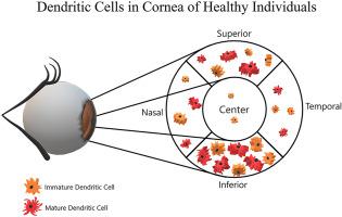
Central corneas had lower DC density (40.83 ± 5.14 cells/mm2; mean ± SEM) as compared to peripheral corneas (75.42 ± 2.67 cells/mm2, p < 0.0001). Inferior and superior zones demonstrated higher DC density (105.01 ± 7.12 and 90.62 ± 4.62 cells/mm2) compared to the nasal and temporal zones (59.93 ± 3.42 and 51.77 ± 2.98 cells/mm2, p < 0.0001). Similarly, lower DC size, field and number of dendrites were observed in the central as compared to the average peripheral cornea (p < 0.0001), with highest values in the inferior zone (p < 0.001 for all, except p < 0.05 for number of dendrites in superior zone). DC parameters did not correlate with age or gender. Inter-observer reliability was 0.987 for DC density and 0.771–0.922 for morphology.
Conclusion
In healthy individuals, the peripheral cornea demonstrates higher DC density and larger morphology compared to the center, with highest values in the inferior zone. We provide the largest normative cohort for sub-stratified DC density and morphology, which can be used in future clinical trials to compare differential changes in diseased states. Furthermore, as DC parameters in the peripheral zones are dissimilar, random sampling of peripheral cornea may be inaccurate.
中文翻译:
使用体内共聚焦显微镜观察健康受试者外周角膜中树突状细胞的密度和分布
目的
使用体内共聚焦显微镜 (IVCM) 在大型健康队列中建立中央和周边角膜的树突状细胞 (DC) 密度和形态学参数。
方法
在 85 名健康志愿者(n = 85 只眼)中进行了一项前瞻性、横断面、观察性研究。分析角膜中心和四个周边区域的 IVCM 图像的 DC 密度和形态,以比较平均值和评估相关性(p < 0.05 具有统计学意义)。
结果
与周边角膜(75.42 ± 2.67 个细胞/mm 2,p < 0.0001)相比,中央角膜具有较低的 DC 密度(40.83 ± 5.14 个细胞/mm 2 ;平均值 ± SEM)。与鼻区和颞区(59.93 ± 3.42 和 51.77 ± 2.98 细胞/mm 2,p < 0.0001)相比,下区和上区表现出更高的 DC 密度(105.01 ± 7.12 和 90.62 ± 4.62 个细胞/mm 2 )。类似地,与平均外周角膜相比,在中央观察到较低的 DC 大小、视野和树突数量 (p < 0.0001),下部区域的值最高 (p < 0.001,除了 p < 0.05 的数量优越区的树突)。DC 参数与年龄或性别无关。DC 密度的观察者间可靠性为 0.987,形态学为 0.771–0.922。
结论
在健康个体中,与中心相比,周边角膜表现出更高的 DC 密度和更大的形态,在下层区域具有最高值。我们为亚分层 DC 密度和形态提供了最大的规范队列,可用于未来的临床试验以比较疾病状态的差异变化。此外,由于周边区域的 DC 参数不同,周边角膜的随机采样可能不准确。


 京公网安备 11010802027423号