Skeletal Radiology ( IF 2.1 ) Pub Date : 2022-08-09 , DOI: 10.1007/s00256-022-04147-w Hwan-Hee Lee 1 , Yeon Soo Lee 2 , Jong Ok Kim 3
|
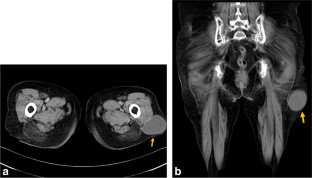
The risk of tuberculosis (TB) increases in immunocompromised patients. Multiple myeloma is considered a risk factor for TB and myeloma patients with TB have a higher mortality rate than those without TB. Herein, we report a case of concomitant TB of the iliotibial band mimicking a soft tissue tumor and tuberculous trochanteric bursitis in a patient with multiple myeloma. In this article, the characteristic magnetic resonance imaging (MRI) findings were low T2 signals in the cystic fluid lesion, a dark T2 signal rim, and peripheral rim enhancement. These results could help differentiate TB of the iliotibial band and trochanteric bursitis from other pathologies. If the abovementioned findings were observed in immunocompromised patients, extrapulmonary TB may be expected even if chest radiographs are normal.
中文翻译:
伴发类似软组织肿瘤的髂胫束结核和结核性转子滑囊炎:一例强调磁共振成像结果的病例报告
免疫功能低下的患者患结核病 (TB) 的风险会增加。多发性骨髓瘤被认为是结核病的危险因素,患有结核病的骨髓瘤患者的死亡率高于未患结核病的患者。在此,我们报告了一例多发性骨髓瘤患者同时患有模拟软组织肿瘤和结核性转子滑囊炎的髂胫束结核病例。在这篇文章中,特征性磁共振成像 (MRI) 发现是囊液病变中的低 T2 信号、暗 T2 信号边缘和周边边缘增强。这些结果可能有助于将髂胫束结核和转子滑囊炎与其他病症区分开来。如果在免疫功能低下的患者中观察到上述发现,即使胸片正常,也可能会发生肺外结核。


 京公网安备 11010802027423号