当前位置:
X-MOL 学术
›
Biotechnol. Bioeng.
›
论文详情
Our official English website, www.x-mol.net, welcomes your
feedback! (Note: you will need to create a separate account there.)
Spheroid culture for chondrocytes triggers the initial stage of endochondral ossification
Biotechnology and Bioengineering ( IF 3.5 ) Pub Date : 2022-08-03 , DOI: 10.1002/bit.28203 Jeonghyun Kim 1 , Kosei Tomida 1 , Takeo Matsumoto 1 , Taiji Adachi 2
Biotechnology and Bioengineering ( IF 3.5 ) Pub Date : 2022-08-03 , DOI: 10.1002/bit.28203 Jeonghyun Kim 1 , Kosei Tomida 1 , Takeo Matsumoto 1 , Taiji Adachi 2
Affiliation
|
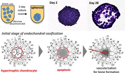
Endochondral ossification is the process of bone formation derived from growing cartilage duringskeletal development. In previous studies, we provoked the osteocyte differentiation of osteoblast precursor cells under a three-dimensional (3D) culture model. To recapitulate the endochondral ossification, the present study utilized the self-organized scaffold-free spheroid model reconstructed by pre-chondrocyte cells. Within 2-day cultivation in the absence of the chemically induced chondrogenesis supplements, the chondrocyte marker was greatly expressed in the inner region of the spheroid, whereas the hypertrophic chondrocyte marker was strongly detected in the surface region of the spheroid. Notably, we found out that the gene expression levels of osteocyte markers were also greatly upregulated compared to the conventional 2D monolayer. Moreover, after long-term cultivation for 28 days, it induced morphological changes in the spheroid, such as cellular hypertrophy and death. In this study, in order to recapitulate the initial stage of the endochondral ossification, we highlighted the potentials of the 3D culture method to drive the hypertrophic chondrocyte differentiation of the pre-chondrocyte cells.
中文翻译:

软骨细胞的球体培养触发了软骨内骨化的初始阶段
软骨内骨化是在骨骼发育过程中由生长的软骨衍生的骨形成过程。在以前的研究中,我们在三维 (3D) 培养模型下激发了成骨细胞前体细胞的骨细胞分化。为了概括软骨内骨化,本研究利用由前软骨细胞重建的自组织无支架球体模型。在没有化学诱导的软骨形成补充剂的情况下的 2 天培养中,软骨细胞标志物在球体的内部区域大量表达,而在球体的表面区域强烈检测到肥大软骨细胞标志物。值得注意的是,我们发现与传统的二维单层相比,骨细胞标志物的基因表达水平也大大上调。而且,长期培养28天后,诱导球体发生细胞肥大、死亡等形态变化。在这项研究中,为了概括软骨内骨化的初始阶段,我们强调了 3D 培养方法驱动前软骨细胞肥大软骨细胞分化的潜力。
更新日期:2022-08-03
中文翻译:
软骨细胞的球体培养触发了软骨内骨化的初始阶段
软骨内骨化是在骨骼发育过程中由生长的软骨衍生的骨形成过程。在以前的研究中,我们在三维 (3D) 培养模型下激发了成骨细胞前体细胞的骨细胞分化。为了概括软骨内骨化,本研究利用由前软骨细胞重建的自组织无支架球体模型。在没有化学诱导的软骨形成补充剂的情况下的 2 天培养中,软骨细胞标志物在球体的内部区域大量表达,而在球体的表面区域强烈检测到肥大软骨细胞标志物。值得注意的是,我们发现与传统的二维单层相比,骨细胞标志物的基因表达水平也大大上调。而且,长期培养28天后,诱导球体发生细胞肥大、死亡等形态变化。在这项研究中,为了概括软骨内骨化的初始阶段,我们强调了 3D 培养方法驱动前软骨细胞肥大软骨细胞分化的潜力。


 京公网安备 11010802027423号