Cell Death and Differentiation ( IF 13.7 ) Pub Date : 2022-08-06 , DOI: 10.1038/s41418-022-01046-4 Fei Yao 1, 2, 3 , Jingjie Peng 1, 2, 3 , Endong Zhang 1, 2, 3 , Dan Ji 1, 2, 3 , Zhaolin Gao 1, 2, 3 , Yixiong Tang 1, 2, 3 , Xueyan Yao 1, 2, 3 , Xiaobo Xia 1, 2, 3
|
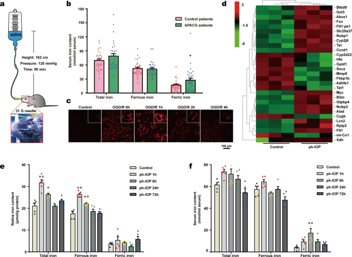
Glaucoma can result in retinal ganglion cell (RGC) death and permanently damaged vision. Pathologically high intraocular pressure (ph-IOP) is the leading cause of damaged vision during glaucoma; however, controlling ph-IOP alone does not entirely prevent the loss of glaucomatous RGCs, and the underlying mechanism remains elusive. In this study, we reported an increase in ferric iron in patients with acute primary angle-closure glaucoma (the most typical glaucoma with ph-IOP damage) compared with the average population by analyzing free iron levels in peripheral serum. Thus, iron metabolism might be involved in regulating the injury of RGCs under ph-IOP. In vitro and in vivo studies confirmed that ph-IOP led to abnormal accumulation of ferrous iron in cells and retinas at 1–8 h post-injury and elevation of ferric iron in serum at 8 h post-injury. Nuclear receptor coactivator 4 (NCOA4)-mediated degradation of ferritin heavy polypeptide 1(FTH1) is essential to disrupt iron metabolism in the retina after ph-IOP injury. Furthermore, knockdown of Ncoa4 in vivo inhibited FTH1 degradation and reduced the retinal ferrous iron level. Elevated ferrous iron induced by ph-IOP led to a marked accumulation of pro-ferroptotic factors (lipid peroxidation and acyl CoA synthetase long-chain family member 4) and a depletion of anti-ferroptotic factors (glutathione, glutathione peroxidase 4, and nicotinamide adenine dinucleotide phosphate). These biochemical changes resulted in RGC ferroptosis. Deferiprone can pass through the blood-retinal barrier after oral administration and chelated abnormally elevated ferrous iron in the retina after ph-IOP injury, thus inhibiting RGC ferroptosis and protecting visual function. In conclusion, this study revealed the role of NCOA4-FTH1-mediated disturbance of iron metabolism and ferroptosis in RGCs during glaucoma. We demonstrate the protective effect of Deferiprone on RGCs via inhibition of ferroptosis, providing a research direction to understand and treat glaucoma via the iron homeostasis and ferroptosis pathways.
中文翻译:
病理性高眼压扰乱正常的铁稳态并导致青光眼视网膜神经节细胞铁死亡
青光眼可导致视网膜神经节细胞 (RGC) 死亡和永久性视力受损。病理性高眼压 (ph-IOP) 是青光眼期间视力受损的主要原因;然而,单独控制 ph-IOP 并不能完全防止青光眼 RGC 的丢失,并且潜在的机制仍然难以捉摸。在这项研究中,我们通过分析外周血清中的游离铁水平,报告了与普通人群相比,急性原发性闭角型青光眼(最典型的伴有 ph-IOP 损伤的青光眼)患者的三价铁含量增加。因此,铁代谢可能参与调节 ph-IOP 下 RGC 的损伤。体外和体内研究证实,ph-IOP 导致损伤后 1-8 小时细胞和视网膜中亚铁的异常积累,以及损伤后 8 小时血清中三价铁的升高。核受体共激活因子 4 (NCOA4) 介导的铁蛋白重多肽 1 (FTH1) 降解对于破坏 ph-IOP 损伤后视网膜中的铁代谢至关重要。此外,击倒Ncoa4在体内抑制 FTH1 降解并降低视网膜亚铁水平。ph-IOP 诱导的亚铁离子升高导致促铁死亡因子(脂质过氧化和酰基辅酶 A 合成酶长链家族成员 4)显着积累和抗铁死亡因子(谷胱甘肽、谷胱甘肽过氧化物酶 4 和烟酰胺腺嘌呤)的消耗磷酸二核苷酸)。这些生化变化导致 RGC 铁死亡。Deferiprone 口服后可通过血视网膜屏障,螯合 ph-IOP 损伤后视网膜中异常升高的亚铁离子,从而抑制 RGC 铁死亡,保护视功能。总之,本研究揭示了青光眼期间 NCOA4-FTH1 介导的 RGC 铁代谢紊乱和铁死亡的作用。通过抑制铁死亡,为通过铁稳态和铁死亡途径了解和治疗青光眼提供了一个研究方向。


 京公网安备 11010802027423号