Our official English website, www.x-mol.net, welcomes your
feedback! (Note: you will need to create a separate account there.)
Raman spectroscopy for the differentiation of Enterobacteriaceae: a comparison of two methods
Analyst ( IF 3.6 ) Pub Date : 2022-08-03 , DOI: 10.1039/d2an00822j Amir Nakar 1, 2, 3 , Annette Wagenhaus 2 , Petra Rösch 2, 3 , Jürgen Popp 1, 2, 3
Analyst ( IF 3.6 ) Pub Date : 2022-08-03 , DOI: 10.1039/d2an00822j Amir Nakar 1, 2, 3 , Annette Wagenhaus 2 , Petra Rösch 2, 3 , Jürgen Popp 1, 2, 3
Affiliation

|
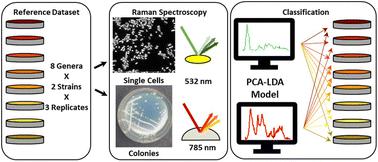
Enterobacteriaceae are the leading cause of urinary tract infections, and include pathogens such as E. coli, K. pneumoniae and P. mirabilis. Due to their similarity, the correct identification of these pathogens is difficult and time-consuming. Raman spectroscopy has been demonstrated extensively as a tool for rapid microbiological differentiation. However, for pathogenic Enterobacteriaceae the application of Raman spectroscopy has been particularly challenging. In this study, two promising methods for Raman-based microbiological diagnostics were compared for differentiating Enterobacteriaceae. Spectra were collected from single-cells with Raman microspectroscopy and from colonies on agar with an NIR Raman fiber-probe. A comprehensive dataset of spectra from 8 different, clinically relevant, genera was collected. Visually, the spectra obtained from both methods presented little difference between the genera. For classification, single cell analysis yielded limited results, while the fiber-probe spectra enabled perfect classification of all 16 isolates. Moreover, the model was validated on new replicates and 15/16 strains were correctly identified (94% overall accuracy). This is the first study to focus on the closely related Enterobacteriaceae, who have previously been avoided or differentiated poorly. It shows how, with the correct spectroscopic setup, even challenging questions in clinical microbiology can be resolved with Raman spectroscopy, highlighting the method's potential for improving patient care.
中文翻译:

区分肠杆菌科细菌的拉曼光谱:两种方法的比较
肠杆菌科细菌是尿路感染的主要原因,包括大肠杆菌、肺炎克雷伯菌和奇异变形杆菌等病原体。由于它们的相似性,正确识别这些病原体既困难又耗时。拉曼光谱已被广泛证明是一种快速微生物分化的工具。然而,对于致病性肠杆菌科细菌,拉曼光谱的应用尤其具有挑战性。在这项研究中,比较了两种有前景的基于拉曼的微生物诊断方法,用于区分肠杆菌科。用拉曼显微光谱法从单细胞收集光谱,用 NIR 拉曼纤维探针从琼脂上的菌落收集光谱。收集了来自 8 个不同的临床相关属的光谱综合数据集。从视觉上看,从两种方法获得的光谱在属之间几乎没有差异。对于分类,单细胞分析产生的结果有限,而光纤探针光谱能够对所有 16 个分离株进行完美分类。此外,该模型在新的复制品上得到了验证,15/16 株被正确识别(94% 的总体准确度)。这是第一项关注密切相关的肠杆菌科的研究,这些肠杆菌科以前被避免或分化不佳。它展示了如何通过正确的光谱设置,即使是临床微生物学中具有挑战性的问题也可以通过拉曼光谱来解决,突出了该方法在改善患者护理方面的潜力。以前被避免或区分不佳的人。它展示了如何通过正确的光谱设置,即使是临床微生物学中具有挑战性的问题也可以通过拉曼光谱来解决,突出了该方法在改善患者护理方面的潜力。以前被避免或区分不佳的人。它展示了如何通过正确的光谱设置,即使是临床微生物学中具有挑战性的问题也可以通过拉曼光谱来解决,突出了该方法在改善患者护理方面的潜力。
更新日期:2022-08-05
中文翻译:

区分肠杆菌科细菌的拉曼光谱:两种方法的比较
肠杆菌科细菌是尿路感染的主要原因,包括大肠杆菌、肺炎克雷伯菌和奇异变形杆菌等病原体。由于它们的相似性,正确识别这些病原体既困难又耗时。拉曼光谱已被广泛证明是一种快速微生物分化的工具。然而,对于致病性肠杆菌科细菌,拉曼光谱的应用尤其具有挑战性。在这项研究中,比较了两种有前景的基于拉曼的微生物诊断方法,用于区分肠杆菌科。用拉曼显微光谱法从单细胞收集光谱,用 NIR 拉曼纤维探针从琼脂上的菌落收集光谱。收集了来自 8 个不同的临床相关属的光谱综合数据集。从视觉上看,从两种方法获得的光谱在属之间几乎没有差异。对于分类,单细胞分析产生的结果有限,而光纤探针光谱能够对所有 16 个分离株进行完美分类。此外,该模型在新的复制品上得到了验证,15/16 株被正确识别(94% 的总体准确度)。这是第一项关注密切相关的肠杆菌科的研究,这些肠杆菌科以前被避免或分化不佳。它展示了如何通过正确的光谱设置,即使是临床微生物学中具有挑战性的问题也可以通过拉曼光谱来解决,突出了该方法在改善患者护理方面的潜力。以前被避免或区分不佳的人。它展示了如何通过正确的光谱设置,即使是临床微生物学中具有挑战性的问题也可以通过拉曼光谱来解决,突出了该方法在改善患者护理方面的潜力。以前被避免或区分不佳的人。它展示了如何通过正确的光谱设置,即使是临床微生物学中具有挑战性的问题也可以通过拉曼光谱来解决,突出了该方法在改善患者护理方面的潜力。


 京公网安备 11010802027423号