Medical Image Analysis ( IF 10.7 ) Pub Date : 2022-07-29 , DOI: 10.1016/j.media.2022.102557 Lisa Kausch 1 , Sarina Thomas 2 , Holger Kunze 3 , Tobias Norajitra 4 , André Klein 2 , Leonardo Ayala 5 , Jan El Barbari 6 , Eric Mandelka 6 , Maxim Privalov 6 , Sven Vetter 6 , Andreas Mahnken 7 , Lena Maier-Hein 8 , Klaus Maier-Hein 4
|
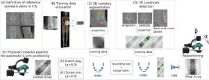
Fluoroscopy-guided trauma and orthopedic surgeries involve the repeated acquisition of correct anatomy-specific standard projections for guidance, monitoring, and evaluating the surgical result. C-arm positioning is usually performed by hand, involving repeated or even continuous fluoroscopy at a cost of radiation exposure and time. We propose to automate this procedure and estimate the pose update for C-arm repositioning directly from a first X-ray without the need for a patient-specific computed tomography scan (CT) or additional technical equipment. Our method is trained on digitally reconstructed radiographs (DRRs) which uniquely provide ground truth labels for an arbitrary number of training examples. The simulated images are complemented with automatically generated segmentations, landmarks, and with simulated k-wires and screws. To successfully achieve a transfer from simulated to real X-rays, and also to increase the interpretability of results, the pipeline was designed to closely reflect the actual clinical decision-making process followed by spinal neurosurgeons. It explicitly incorporates steps such as region-of-interest (ROI) localization, detection of relevant and view-independent landmarks, and subsequent pose regression. The method was validated on a large human cadaver study simulating a real clinical scenario, including k-wires and screws. The proposed procedure obtained superior C-arm positioning accuracy of average improvement (), robustness, and generalization capabilities compared to the state-of-the-art direct pose regression framework.
中文翻译:
脊柱植入物植入期间用于标准投影的 C 形臂定位
荧光镜引导的创伤和骨科手术涉及重复获取正确的解剖学特定标准投影,以指导、监测和评估手术结果。C 形臂定位通常由手工执行,涉及重复甚至连续透视,以辐射暴露和时间为代价。我们建议自动执行此过程并直接从第一张 X 射线估计 C 臂重新定位的姿势更新,而无需针对患者的计算机断层扫描 (CT) 或其他技术设备。我们的方法是在数字重建射线照片 (DRR) 上进行训练的,它为任意数量的训练示例提供了唯一的地面真实标签。模拟图像与自动生成的分割、地标以及模拟克氏针和螺钉相辅相成。为了成功实现从模拟 X 射线到真实 X 射线的转换,并提高结果的可解释性,管道的设计旨在密切反映脊柱神经外科医生遵循的实际临床决策过程。它明确包含感兴趣区域 (ROI) 定位、相关和视图独立地标检测以及后续姿态回归等步骤。该方法在模拟真实临床场景(包括克氏针和螺钉)的大型人体尸体研究中得到验证。所提出的程序获得了优越的 C 臂定位精度 它明确包含感兴趣区域 (ROI) 定位、相关和视图独立地标检测以及后续姿态回归等步骤。该方法在模拟真实临床场景(包括克氏针和螺钉)的大型人体尸体研究中得到验证。所提出的程序获得了优越的 C 臂定位精度 它明确包含感兴趣区域 (ROI) 定位、相关和视图独立地标检测以及后续姿态回归等步骤。该方法在模拟真实临床场景(包括克氏针和螺钉)的大型人体尸体研究中得到验证。所提出的程序获得了优越的 C 臂定位精度平均改善()、鲁棒性和泛化能力与最先进的直接姿态回归框架相比。


 京公网安备 11010802027423号