European Journal of Nuclear Medicine and Molecular Imaging ( IF 8.6 ) Pub Date : 2022-07-29 , DOI: 10.1007/s00259-022-05901-x René Hosch 1, 2 , Manuel Weber 3 , Miriam Sraieb 3 , Nils Flaschel 1, 2 , Johannes Haubold 1 , Moon-Sung Kim 1, 2 , Lale Umutlu 1 , Jens Kleesiek 2 , Ken Herrmann 3 , Felix Nensa 1, 2 , Christoph Rischpler 3 , Sven Koitka 1, 2 , Robert Seifert 3, 4 , David Kersting 3
|
Purpose
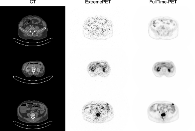
Both digital positron emission tomography (PET) detector technologies and artificial intelligence based image post-reconstruction methods allow to reduce the PET acquisition time while maintaining diagnostic quality. The aim of this study was to acquire ultra-low-count fluorodeoxyglucose (FDG) ExtremePET images on a digital PET/computed tomography (CT) scanner at an acquisition time comparable to a CT scan and to generate synthetic full-dose PET images using an artificial neural network.
Methods
This is a prospective, single-arm, single-center phase I/II imaging study. A total of 587 patients were included. For each patient, a standard and an ultra-low-count FDG PET/CT scan (whole-body acquisition time about 30 s) were acquired. A modified pix2pixHD deep-learning network was trained employing 387 data sets as training and 200 as test cohort. Three models (PET-only and PET/CT with or without group convolution) were compared. Detectability and quantification were evaluated.
Results
The PET/CT input model with group convolution performed best regarding lesion signal recovery and was selected for detailed evaluation. Synthetic PET images were of high visual image quality; mean absolute lesion SUVmax (maximum standardized uptake value) difference was 1.5. Patient-based sensitivity and specificity for lesion detection were 79% and 100%, respectively. Not-detected lesions were of lower tracer uptake and lesion volume. In a matched-pair comparison, patient-based (lesion-based) detection rate was 89% (78%) for PERCIST (PET response criteria in solid tumors)-measurable and 36% (22%) for non PERCIST-measurable lesions.
Conclusion
Lesion detectability and lesion quantification were promising in the context of extremely fast acquisition times. Possible application scenarios might include re-staging of late-stage cancer patients, in whom assessment of total tumor burden can be of higher relevance than detailed evaluation of small and low-uptake lesions.
中文翻译:
人工智能引导增强数字PET:扫描速度与CT一样快?
目的
数字正电子发射断层扫描 (PET) 检测器技术和基于人工智能的图像后重建方法都可以减少 PET 采集时间,同时保持诊断质量。本研究的目的是在数字 PET/计算机断层扫描 (CT) 扫描仪上以与 CT 扫描相当的采集时间获取超低计数氟脱氧葡萄糖 (FDG) ExtremePET 图像,并使用人工神经网络。
方法
这是一项前瞻性、单臂、单中心 I/II 期影像学研究。总共包括 587 名患者。对每位患者进行标准和超低计数 FDG PET/CT 扫描(全身采集时间约 30 秒)。修改后的 pix2pixHD 深度学习网络使用 387 个数据集作为训练数据集,200 个数据集作为测试队列进行训练。比较了三种模型(仅 PET 和有或没有组卷积的 PET/CT)。评估了可检测性和量化。
结果
具有组卷积的 PET/CT 输入模型在病变信号恢复方面表现最佳,并被选中进行详细评估。合成PET图像视觉图像质量高;平均绝对病变 SUV最大值(最大标准化摄取值)差异为 1.5。基于患者的病变检测敏感性和特异性分别为 79% 和 100%。未检测到的病变具有较低的示踪剂摄取和病变体积。在配对比较中,PERCIST(实体瘤中的 PET 反应标准)可测量的基于患者(基于病灶)的检出率为 89%(78%),非 PERCIST 可测量病灶的检出率为 36%(22%)。
结论
在极快的采集时间的背景下,病变可检测性和病变量化是有前途的。可能的应用场景可能包括晚期癌症患者的重新分期,在这些患者中,对总肿瘤负荷的评估可能比对小和低摄取病变的详细评估具有更高的相关性。


 京公网安备 11010802027423号