Acta Neuropathologica ( IF 9.3 ) Pub Date : 2022-07-27 , DOI: 10.1007/s00401-022-02469-6 Antonio Boza-Serrano 1, 2, 3 , Agathe Vrillon 4, 5 , Karolina Minta 6 , Agnes Paulus 1, 7 , Lluís Camprubí-Ferrer 1 , Megg Garcia 1, 8 , Ulf Andreasson 6, 9 , Anna Antonell 2 , Malin Wennström 10 , Gunnar Gouras 8 , Julien Dumurgier 4, 5 , Emmanuel Cognat 4, 5 , Laura Molina-Porcel 2, 11 , Mircea Balasa 2 , Javier Vitorica 3, 12 , Raquel Sánchez-Valle 2 , Claire Paquet 4, 5 , Jose Luis Venero 3 , Kaj Blennow 6, 9 , Tomas Deierborg 1
|
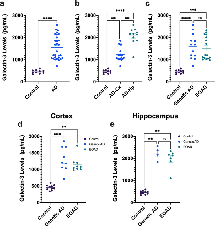
Galectin-3 (Gal-3) is a beta-galactosidase binding protein involved in microglial activation in the central nervous system (CNS). We previously demonstrated the crucial deleterious role of Gal-3 in microglial activation in Alzheimer’s disease (AD). Under AD conditions, Gal-3 is primarily expressed by microglial cells clustered around Aβ plaques in both human and mouse brain, and knocking out Gal-3 reduces AD pathology in AD-model mice. To further unravel the importance of Gal-3-associated inflammation in AD, we aimed to investigate the Gal-3 inflammatory response in the AD continuum. First, we measured Gal-3 levels in neocortical and hippocampal tissue from early-onset AD patients, including genetic and sporadic cases. We found that Gal-3 levels were significantly higher in both cortex and hippocampus in AD subjects. Immunohistochemistry revealed that Gal-3+ microglial cells were associated with amyloid plaques of a larger size and more irregular shape and with neurons containing tau-inclusions. We then analyzed the levels of Gal-3 in cerebrospinal fluid (CSF) from AD patients (n = 119) compared to control individuals (n = 36). CSF Gal-3 levels were elevated in AD patients compared to controls and more strongly correlated with tau (p-Tau181 and t-tau) and synaptic markers (GAP-43 and neurogranin) than with amyloid-β. Lastly, principal component analysis (PCA) of AD biomarkers revealed that CSF Gal-3 clustered and associated with other CSF neuroinflammatory markers, including sTREM-2, GFAP, and YKL-40. This neuroinflammatory component was more highly expressed in the CSF from amyloid-β positive (A+), CSF p-Tau181 positive (T+), and biomarker neurodegeneration positive/negative (N+/−) (A + T + N+/−) groups compared to the A + T−N− group. Overall, Gal-3 stands out as a key pathological biomarker of AD pathology that is measurable in CSF and, therefore, a potential target for disease-modifying therapies involving the neuroinflammatory response.
中文翻译:
Galectin-3 在 CSF 中升高,并与阿尔茨海默病脑组织中的 Aβ 沉积物和 tau 聚集体相关
Galectin-3 (Gal-3) 是一种 β-半乳糖苷酶结合蛋白,参与中枢神经系统 (CNS) 中小胶质细胞的激活。我们之前证明了 Gal-3 在阿尔茨海默病 (AD) 小胶质细胞激活中的关键有害作用。在 AD 条件下,Gal-3 主要由聚集在人和小鼠大脑中 Aβ 斑块周围的小胶质细胞表达,敲除 Gal-3 可减少 AD 模型小鼠的 AD 病理。为了进一步阐明 Gal-3 相关炎症在 AD 中的重要性,我们旨在研究 AD 连续体中的 Gal-3 炎症反应。首先,我们测量了早发性 AD 患者(包括遗传性和散发性病例)的新皮质和海马组织中的 Gal-3 水平。我们发现 Gal-3 水平在 AD 受试者的皮质和海马体中显着更高。免疫组织化学表明,Gal-3+ 小胶质细胞与更大尺寸和更不规则形状的淀粉样斑块以及含有 tau 包涵体的神经元相关。然后我们分析了 AD 患者脑脊液 (CSF) 中 Gal-3 的水平(n = 119) 与对照个体相比 ( n = 36). 与对照组相比,AD 患者的 CSF Gal-3 水平升高,并且与 tau(p-Tau181 和 t-tau)和突触标志物(GAP-43 和神经粒蛋白)的相关性比与淀粉样蛋白-β 的相关性更强。最后,AD 生物标志物的主成分分析 (PCA) 显示 CSF Gal-3 聚集并与其他 CSF 神经炎症标志物相关,包括 sTREM-2、GFAP 和 YKL-40。相比之下,这种神经炎症成分在淀粉样蛋白-β 阳性 (A+)、CSF p-Tau181 阳性 (T+) 和生物标志物神经变性阳性/阴性 (N+/-) (A + T + N+/-) 组的 CSF 中表达更高到 A + T−N− 组。总体而言,Gal-3 作为 AD 病理学的关键病理生物标志物脱颖而出,可在 CSF 中测量,因此,它是涉及神经炎症反应的疾病缓解疗法的潜在靶标。


 京公网安备 11010802027423号