Journal of Materials Science & Technology ( IF 11.2 ) Pub Date : 2022-07-24 , DOI: 10.1016/j.jmst.2022.06.036 Bo Chen , Zhengjie Lin , Qimanguli Saiding , Yongcan Huang , Yi Sun , Xinyun Zhai , Ziyu Ning , Hai Liang , Wei Qiao , Bingsheng Yu , Kelvin W.K. Yeung , Jie Shen
|
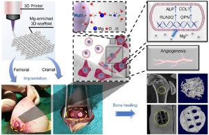
The healing of critical-sized bone defects (CSD) remains a challenge in orthopaedic medicine. In recent years, scaffolds with sophisticated microstructures fabricated by the emerging three-dimensional (3D) printing technology have lighted up the treatment of the CSD due to the elaborate microenvironments and support they may build. Here, we established a magnesium oxide-reinforced 3D-printed biocomposite scaffold to investigate the effect of magnesium-enriched 3D microenvironment on CSD repairing. The composite was prepared using a biodegradable polymer matrix, polycaprolactone (PCL), and the dispersion phase, magnesium oxide (MgO). With the appropriate surface treatment by saline coupling agent, the MgO dispersed homogeneously in the polymer matrix, leading to enhanced mechanical performance and steady release of magnesium ion (Mg2+) for superior cytocompatibility, higher cell viability, advanced osteogenic differentiation, and cell mineralization capabilities in comparison with the pure PCL. The in-vivo femoral implantation and critical-sized cranial bone defect studies demonstrated the importance of the 3D magnesium microenvironment, as a scaffold that released appropriate Mg2+ exhibited remarkably increased bone volume, enhanced angiogenesis, and almost recovered CSD after 8-week implantation. Overall, this study suggests that the magnesium-enriched 3D scaffold is a potential candidate for the treatment of CSD in a cell-free therapeutic approach.
中文翻译:
通过具有改善的成骨和血管生成特性的氧化镁增强的 3D 支架增强临界大小的骨缺损再生
临界尺寸骨缺损 (CSD) 的愈合仍然是骨科医学的一项挑战。近年来,通过新兴的 3D (3D) 打印技术制造的具有复杂微结构的支架由于其可能构建的精细微环境和支持而照亮了 CSD 的治疗。在这里,我们建立了一种氧化镁增强的 3D 打印生物复合材料支架,以研究富含镁的 3D 微环境对 CSD 修复的影响。该复合材料是使用可生物降解的聚合物基质聚己内酯 (PCL) 和分散相氧化镁 (MgO) 制备的。通过盐水偶联剂进行适当的表面处理,MgO 均匀分散在聚合物基体中,从而提高了机械性能并稳定释放了镁离子(Mg2+ ) 与纯 PCL 相比,具有出色的细胞相容性、更高的细胞活力、先进的成骨分化和细胞矿化能力。体内股骨植入和临界大小颅骨缺损研究证明了 3D 镁微环境的重要性,作为释放适当 Mg 2+ 的支架,在植入8 周后骨量显着增加,血管生成增强,CSD 几乎恢复. 总体而言,这项研究表明,富含镁的 3D 支架是在无细胞治疗方法中治疗 CSD 的潜在候选者。


 京公网安备 11010802027423号