当前位置:
X-MOL 学术
›
Microsc. Res. Tech.
›
论文详情
Our official English website, www.x-mol.net, welcomes your feedback! (Note: you will need to create a separate account there.)
Histomorphology and ultrastructure of the proventriculus of the broad breasted white turkey (Meleagris gallopavo, Linnaeus 1758)
Microscopy Research and Technique ( IF 2.5 ) Pub Date : 2022-07-22 , DOI: 10.1002/jemt.24203 Fatma A Madkour 1 , Ramadan M Kandyel 2
Microscopy Research and Technique ( IF 2.5 ) Pub Date : 2022-07-22 , DOI: 10.1002/jemt.24203 Fatma A Madkour 1 , Ramadan M Kandyel 2
Affiliation
|
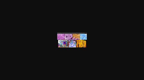
This study aims to examine the functional morphology of the proventriculus of the broad breasted white turkey (BBWT) (Meleagris gallopavo, Linnaeus 1758) using gross anatomy, light microscopy, gross/histomorphometric analysis, and scanning and transmission electron microscopy. The proventriculus was characterized internally by many elevated papillae with a mound, leafy flower, and lotus flower-shapes. Each papilla was enclosed by concentrically organized mucosal folds with distinct or indistinct proventricular gland openings on its top. Longitudinal folds and grooves at the junction of the proventriculus with the esophagus exhibited various sized and shaped openings of esophageal glands with irregular outlines. Histologically, the surface epithelium of the proventriculus was covered by a thin layer of cuticle, particularly evident at its junction with the gizzard. The lamina epithelialis and propria, and secretory units of proventricular lobules were infiltrated by aggregations of lymphocytes and lymphoid follicles (nodules). Variably shaped glandular lobules of proventricular glands occupied the submucosa, surrounded by thin strands of smooth muscle fibers derived from muscularis mucosa. Triangular, cuboidal, or columnar-shaped secretory oxyntico-peptic cells lined the secretory units. Many telocytes (pyramidal or fusiform-shaped cell bodies with lengthy telopodes) were observed in interstitial tissue. Further, two types of argyrophilic endocrine cells were identified within the glandular epithelium. The morphology of the M. gallopavo proventriculus reflects its dietary habits and behavior.
中文翻译:

宽胸白火鸡胃腺的组织形态学和超微结构 (Meleagris gallopavo, Linnaeus 1758)
本研究旨在检查宽胸白火鸡 (BBWT) ( Meleagris gallopavo ) 胃腺的功能形态。, Linnaeus 1758) 使用大体解剖、光学显微镜、大体/组织形态学分析以及扫描和透射电子显微镜。腺胃的内部特征是许多隆起的乳头状突起,呈丘状、叶状花状和莲花状。每个乳头被同心组织的粘膜皱襞包围,顶部有明显或不明显的前室腺开口。腺胃与食管交界处的纵向皱襞和凹槽显示出大小不一、形状不一的食管腺开口,轮廓不规则。组织学上,腺胃的表面上皮被一层薄薄的角质层覆盖,在其与砂囊的交界处尤其明显。上皮层和固有层,前室小叶和分泌单位被淋巴细胞和淋巴滤泡(结节)的聚集浸润。前室腺的不同形状的腺体小叶占据粘膜下层,被来自粘膜肌层的平滑肌纤维细丝包围。三角形、立方形或柱状分泌泌酸-消化细胞排列在分泌单元上。在间质组织中观察到许多端细胞(锥体状或梭形细胞体,端部较长)。此外,在腺上皮内鉴定出两种类型的嗜银内分泌细胞。的形态学 立方形或柱状分泌泌酸消化细胞排列在分泌单位内。在间质组织中观察到许多端细胞(锥体状或梭形细胞体,端部较长)。此外,在腺上皮内鉴定出两种类型的嗜银内分泌细胞。的形态学 立方形或柱状分泌泌酸消化细胞排列在分泌单位内。在间质组织中观察到许多端细胞(锥体状或梭形细胞体,端部较长)。此外,在腺上皮内鉴定出两种类型的嗜银内分泌细胞。的形态学米。Gallopavo Proventriculus 反映了它的饮食习惯和行为。
更新日期:2022-07-22
中文翻译:
宽胸白火鸡胃腺的组织形态学和超微结构 (Meleagris gallopavo, Linnaeus 1758)
本研究旨在检查宽胸白火鸡 (BBWT) ( Meleagris gallopavo ) 胃腺的功能形态。, Linnaeus 1758) 使用大体解剖、光学显微镜、大体/组织形态学分析以及扫描和透射电子显微镜。腺胃的内部特征是许多隆起的乳头状突起,呈丘状、叶状花状和莲花状。每个乳头被同心组织的粘膜皱襞包围,顶部有明显或不明显的前室腺开口。腺胃与食管交界处的纵向皱襞和凹槽显示出大小不一、形状不一的食管腺开口,轮廓不规则。组织学上,腺胃的表面上皮被一层薄薄的角质层覆盖,在其与砂囊的交界处尤其明显。上皮层和固有层,前室小叶和分泌单位被淋巴细胞和淋巴滤泡(结节)的聚集浸润。前室腺的不同形状的腺体小叶占据粘膜下层,被来自粘膜肌层的平滑肌纤维细丝包围。三角形、立方形或柱状分泌泌酸-消化细胞排列在分泌单元上。在间质组织中观察到许多端细胞(锥体状或梭形细胞体,端部较长)。此外,在腺上皮内鉴定出两种类型的嗜银内分泌细胞。的形态学 立方形或柱状分泌泌酸消化细胞排列在分泌单位内。在间质组织中观察到许多端细胞(锥体状或梭形细胞体,端部较长)。此外,在腺上皮内鉴定出两种类型的嗜银内分泌细胞。的形态学 立方形或柱状分泌泌酸消化细胞排列在分泌单位内。在间质组织中观察到许多端细胞(锥体状或梭形细胞体,端部较长)。此外,在腺上皮内鉴定出两种类型的嗜银内分泌细胞。的形态学米。Gallopavo Proventriculus 反映了它的饮食习惯和行为。


 京公网安备 11010802027423号