Acta Neuropathologica ( IF 9.3 ) Pub Date : 2022-07-22 , DOI: 10.1007/s00401-022-02466-9 Evelien Van Schoor 1, 2, 3 , Simona Ospitalieri 1 , Sebastiaan Moonen 1, 3, 4 , Sandra O Tomé 1 , Alicja Ronisz 1 , Orkun Ok 1 , Jochen Weishaupt 5, 6 , Albert C Ludolph 5, 7 , Philip Van Damme 2, 3, 8 , Ludo Van Den Bosch 2, 3 , Dietmar Rudolf Thal 1, 9
|
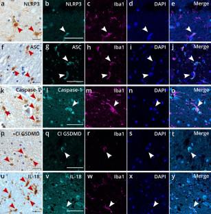
Amyotrophic lateral sclerosis (ALS) is characterized by the degeneration of motor neurons in the motor cortex, brainstem, and spinal cord. Although ALS is considered a motor neuron disorder, neuroinflammation also plays an important role. Recent evidence in ALS disease models indicates activation of the inflammasome and subsequent initiation of pyroptosis, an inflammatory type of cell death. In this study, we determined the expression and distribution of the inflammasome and pyroptosis effector proteins in post-mortem brain and spinal cord from ALS patients (n = 25) and controls (n = 19), as well as in symptomatic and asymptomatic TDP-43A315T transgenic and wild-type mice. Furthermore, we evaluated its correlation with the presence of TDP-43 pathological proteins and neuronal loss. Expression of the NOD-, LRR-, and pyrin domain-containing protein 3 (NLRP3) inflammasome, pyroptosis effector protein cleaved Gasdermin D (GSDMD), and IL-18 was detected in microglia in human ALS motor cortex and spinal cord, indicative of canonical inflammasome-triggered pyroptosis activation. The number of cleaved GSDMD-positive precentral white matter microglia was increased compared to controls and correlated with a decreased neuronal density in human ALS motor cortex. Neither of this was observed in the spinal cord. Similar results were obtained in TDP-43A315T mice, where microglial pyroptosis activation was significantly increased in the motor cortex upon symptom onset, and correlated with neuronal loss. There was no significant correlation with the presence of TDP-43 pathological proteins both in human and mouse tissue. Our findings emphasize the importance of microglial NLRP3 inflammasome-mediated pyroptosis activation for neuronal degeneration in ALS and pave the way for new therapeutic strategies counteracting motor neuron degeneration in ALS by inhibiting microglial inflammasome/pyroptosis activation.
中文翻译:
白质小胶质细胞焦亡激活增加与 ALS 运动皮层神经元丢失有关
肌萎缩侧索硬化症 (ALS) 的特征是运动皮层、脑干和脊髓中的运动神经元退化。尽管 ALS 被认为是一种运动神经元疾病,但神经炎症也起着重要作用。最近在 ALS 疾病模型中的证据表明炎症小体的激活和随后的细胞焦亡(一种炎症类型的细胞死亡)的启动。在这项研究中,我们确定了 ALS 患者 ( n = 25) 和对照组 ( n = 19) 以及有症状和无症状TDP-死后大脑和脊髓中炎性体和焦亡效应蛋白的表达和分布。43 A315T转基因和野生型小鼠。此外,我们评估了它与 TDP-43 病理蛋白的存在和神经元丢失的相关性。在人类 ALS 运动皮层和脊髓的小胶质细胞中检测到 NOD、LRR 和 pyrin 结构域蛋白 3 (NLRP3) 炎性体、焦亡效应蛋白裂解 Gasdermin D (GSDMD) 和 IL-18 的表达,表明典型的炎症小体触发的细胞焦亡激活。与对照组相比,切割的 GSDMD 阳性前中央白质小胶质细胞的数量增加,并且与人类 ALS 运动皮层中神经元密度的降低相关。在脊髓中均未观察到这两种情况。在 TDP-43 A315T中也获得了类似的结果小鼠,其中小胶质细胞焦亡激活在症状发作时在运动皮层中显着增加,并且与神经元损失相关。与人和小鼠组织中 TDP-43 病理蛋白的存在没有显着相关性。我们的研究结果强调了小胶质细胞 NLRP3 炎症小体介导的细胞焦亡激活对 ALS 神经元变性的重要性,并为通过抑制小胶质细胞炎症小体/细胞焦亡激活来对抗 ALS 运动神经元变性的新治疗策略铺平了道路。


 京公网安备 11010802027423号