当前位置:
X-MOL 学术
›
Microsc. Res. Tech.
›
论文详情
Our official English website, www.x-mol.net, welcomes your feedback! (Note: you will need to create a separate account there.)
Multiscale segmentation- and error-guided iterative convolutional neural network for cerebral neuron segmentation in microscopic images
Microscopy Research and Technique ( IF 2.5 ) Pub Date : 2022-07-20 , DOI: 10.1002/jemt.24206 Zhenzhen You 1, 2 , Ming Jiang 3 , Zhenghao Shi 1 , Minghua Zhao 1 , Cheng Shi 1 , Shuangli Du 1 , Anne-Sophie Hérard 2 , Nicolas Souedet 2 , Thierry Delzescaux 2
Microscopy Research and Technique ( IF 2.5 ) Pub Date : 2022-07-20 , DOI: 10.1002/jemt.24206 Zhenzhen You 1, 2 , Ming Jiang 3 , Zhenghao Shi 1 , Minghua Zhao 1 , Cheng Shi 1 , Shuangli Du 1 , Anne-Sophie Hérard 2 , Nicolas Souedet 2 , Thierry Delzescaux 2
Affiliation
|
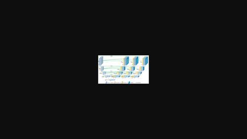
This article uses microscopy images obtained from diverse anatomical regions of macaque brain for neuron semantic segmentation. The complex structure of brain, the large intra-class staining intensity difference within neuron class, the small inter-class staining intensity difference between neuron and tissue class, and the unbalanced dataset increase the difficulty of neuron semantic segmentation. To address this problem, we propose a multiscale segmentation- and error-guided iterative convolutional neural network (MSEG-iCNN) to improve the semantic segmentation performance in major anatomical regions of the macaque brain. After evaluating microscopic images from 17 anatomical regions, the semantic segmentation performance of neurons is improved by 10.6%, 4.0%, 1.5%, and 1.2% compared with Random Forest, FCN-8s, U-Net, and UNet++, respectively. Especially for neurons with brighter staining intensity in the anatomical regions such as lateral geniculate, globus pallidus and hypothalamus, the performance is improved by 66.1%, 23.9%, 11.2%, and 6.7%, respectively. Experiments show that our proposed method can efficiently segment neurons with a wide range of staining intensities. The semantic segmentation results are of great significance and can be further used for neuron instance segmentation, morphological analysis and disease diagnosis. Cell segmentation plays a critical role in extracting cerebral information, such as cell counting, cell morphometry and distribution analysis. Accurate automated neuron segmentation is challenging due to the complex structure of brain, the large intra-class staining intensity difference within neuron class, the small inter-class staining intensity difference between neuron and tissue class, and the unbalanced dataset. The proposed multiscale segmentation- and error-guided iterative convolutional neural network (MSEG-iCNN) improve the segmentation performance in 17 major anatomical regions of the macaque brain.
中文翻译:

用于显微图像中脑神经元分割的多尺度分割和错误引导的迭代卷积神经网络
本文使用从猕猴大脑不同解剖区域获得的显微镜图像进行神经元语义分割。大脑结构复杂,神经元类内类内染色强度差异大,神经元与组织类间染色强度差异小,数据集不平衡增加了神经元语义分割的难度。为了解决这个问题,我们提出了一种多尺度分割和错误引导的迭代卷积神经网络 (MSEG-iCNN),以提高猕猴大脑主要解剖区域的语义分割性能。在评估来自 17 个解剖区域的显微图像后,与随机森林、FCN-8s、U-Net 和 UNet++ 相比,神经元的语义分割性能分别提高了 10.6%、4.0%、1.5% 和 1.2%。尤其是外侧膝状体、苍白球和下丘脑等解剖区域染色强度较亮的神经元,性能分别提高了66.1%、23.9%、11.2%和6.7%。实验表明,我们提出的方法可以有效地分割具有广泛染色强度的神经元。语义分割结果意义重大,可进一步用于神经元实例分割、形态分析和疾病诊断。细胞分割在提取大脑信息方面起着至关重要的作用,例如细胞计数、细胞形态测量和分布分析。由于大脑的复杂结构,神经元类内的大类内染色强度差异,准确的自动神经元分割具有挑战性,神经元和组织类之间的小类间染色强度差异,以及不平衡的数据集。所提出的多尺度分割和错误引导的迭代卷积神经网络 (MSEG-iCNN) 提高了猕猴大脑 17 个主要解剖区域的分割性能。
更新日期:2022-07-20
中文翻译:
用于显微图像中脑神经元分割的多尺度分割和错误引导的迭代卷积神经网络
本文使用从猕猴大脑不同解剖区域获得的显微镜图像进行神经元语义分割。大脑结构复杂,神经元类内类内染色强度差异大,神经元与组织类间染色强度差异小,数据集不平衡增加了神经元语义分割的难度。为了解决这个问题,我们提出了一种多尺度分割和错误引导的迭代卷积神经网络 (MSEG-iCNN),以提高猕猴大脑主要解剖区域的语义分割性能。在评估来自 17 个解剖区域的显微图像后,与随机森林、FCN-8s、U-Net 和 UNet++ 相比,神经元的语义分割性能分别提高了 10.6%、4.0%、1.5% 和 1.2%。尤其是外侧膝状体、苍白球和下丘脑等解剖区域染色强度较亮的神经元,性能分别提高了66.1%、23.9%、11.2%和6.7%。实验表明,我们提出的方法可以有效地分割具有广泛染色强度的神经元。语义分割结果意义重大,可进一步用于神经元实例分割、形态分析和疾病诊断。细胞分割在提取大脑信息方面起着至关重要的作用,例如细胞计数、细胞形态测量和分布分析。由于大脑的复杂结构,神经元类内的大类内染色强度差异,准确的自动神经元分割具有挑战性,神经元和组织类之间的小类间染色强度差异,以及不平衡的数据集。所提出的多尺度分割和错误引导的迭代卷积神经网络 (MSEG-iCNN) 提高了猕猴大脑 17 个主要解剖区域的分割性能。


 京公网安备 11010802027423号