Nature Structural & Molecular Biology ( IF 12.5 ) Pub Date : 2022-07-14 , DOI: 10.1038/s41594-022-00792-w Francesca Vallese 1, 2, 3 , Kookjoo Kim 1, 2, 3 , Laura Y Yen 2 , Jake D Johnston 2, 4 , Alex J Noble 4 , Tito Calì 5, 6, 7 , Oliver Biggs Clarke 1, 2, 3
|
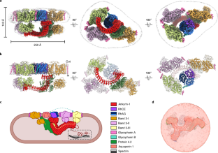
The stability and shape of the erythrocyte membrane is provided by the ankyrin-1 complex, but how it tethers the spectrin–actin cytoskeleton to the lipid bilayer and the nature of its association with the band 3 anion exchanger and the Rhesus glycoproteins remains unknown. Here we present structures of ankyrin-1 complexes purified from human erythrocytes. We reveal the architecture of a core complex of ankyrin-1, the Rhesus proteins RhAG and RhCE, the band 3 anion exchanger, protein 4.2, glycophorin A and glycophorin B. The distinct T-shaped conformation of membrane-bound ankyrin-1 facilitates recognition of RhCE and, unexpectedly, the water channel aquaporin-1. Together, our results uncover the molecular details of ankyrin-1 association with the erythrocyte membrane, and illustrate the mechanism of ankyrin-mediated membrane protein clustering.
中文翻译:
人红细胞 ankyrin-1 复合物的结构
红细胞膜的稳定性和形状由锚蛋白-1 复合物提供,但它如何将血影蛋白-肌动蛋白细胞骨架束缚到脂质双层以及其与带 3 阴离子交换剂和恒河猴糖蛋白的关联性质仍然未知。在这里,我们展示了从人红细胞中纯化的 ankyrin-1 复合物的结构。我们揭示了 ankyrin-1、恒河猴蛋白 RhAG 和 RhCE、带 3 阴离子交换剂、蛋白 4.2、血型糖蛋白 A 和血型糖蛋白 B 的核心复合物的结构。膜结合的 ankyrin-1 独特的 T 形构象有助于识别RhCE 和出人意料的水通道水通道蛋白-1。总之,我们的结果揭示了 ankyrin-1 与红细胞膜关联的分子细节,并说明了锚蛋白介导的膜蛋白聚集的机制。


 京公网安备 11010802027423号