European Journal of Nuclear Medicine and Molecular Imaging ( IF 8.6 ) Pub Date : 2022-07-13 , DOI: 10.1007/s00259-022-05903-9 Yan Wang 1, 2 , Chao Wang 3 , Minzhou Huang 1, 2 , Songbing Qin 4 , Jun Zhao 5 , Shibiao Sang 6 , Meng Zheng 1, 2 , Yicong Bian 1, 2 , Chenrong Huang 1, 2 , Hua Zhang 1, 2 , Lingchuan Guo 7 , Jiwei Jiang 6 , Chun Xu 5 , Na Dai 6 , Yushuang Zheng 7 , Jiajun Han 5 , Min Yang 8 , Tao Xu 3 , Liyan Miao 1, 2
|
Purpose
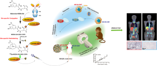
Positron emission tomography (PET) with specific diagnostic probes for quantifying CD8+ T cells has emerged as a powerful technique for monitoring the immune response. However, most CD8+ T cell radiotracers are based on antibodies or antibody fragments, which are slowly cleared from circulation. Herein, we aimed to develop and assess 68 Ga-NODAGA-SNA006 for instant PET (iPET) imaging of CD8+ T cells.
Methods
A novel nanobody without a hexahistidine (His6) tag, SNA006-GSC, was designed, site-specifically conjugated with NODAGA-maleimide and radiolabelled with 68 Ga. The PET imaging profiles of 68 Ga-NODAGA-SNA006 were evaluated in BALB/c MC38-CD8+/CD8− tumour models and cynomolgus monkeys. Three volunteers with lung cancer underwent whole-body PET/CT imaging after 68 Ga-NODAGA-SNA006 administration. The biodistribution, pharmacokinetics and dosimetry of patients were also investigated. In addition, combined with immunohistochemistry (IHC), the quantitative performance of the tracer for monitoring CD8 expression was evaluated in BALB/c MC38-CD8+/CD8− and human subjects.
Results
68 Ga-NODAGA-SNA006 was prepared with RCP > 98% and SA > 100 GBq/μmol. 68 Ga-NODAGA-SNA006 exhibited specific uptake in MC38-CD8+ xenografts tumours, CD8-rich tissues (such as the spleen) in monkeys and CD8+ tumour lesions in patients within 1 h. Fast washout from circulation was observed in three volunteers (t1/2 < 20 min). A preliminary quantitative linear relationship (R2 = 0.9668, p < 0.0001 for xenografts and R2 = 0.7924, p = 0.0013 for lung patients) appeared between 68 Ga-NODAGA-SNA006 uptake and CD8 expression. 68 Ga-NODAGA-SNA006 was well tolerated by all patients.
Conclusion
68 Ga-NODAGA-SNA006 PET imaging can instantly quantify CD8 expression with an ideal safety profile and is expected to be important for dynamically tracking CD8+ T cells and monitoring immune responses for individualised cancer immunotherapy.
Trial registration
NCT05126927 (19 November 2021, retrospectively registered).
中文翻译:
用于 CD8+ T 细胞即时 PET 成像的新型纳米抗体 68 Ga-NODAGA-SNA006 的初步研究
目的
具有用于量化 CD8 + T 细胞的特定诊断探针的正电子发射断层扫描 (PET)已成为监测免疫反应的强大技术。然而,大多数 CD8 + T 细胞放射性示踪剂是基于抗体或抗体片段,它们会慢慢从循环中清除。在此,我们旨在开发和评估用于 CD8 + T 细胞即时 PET (iPET) 成像的68 Ga-NODAGA-SNA006 。
方法
设计了一种没有六组氨酸 (His 6 ) 标签的新型纳米抗体 SNA006-GSC,它与 NODAGA-马来酰亚胺位点特异性结合并用68 Ga 进行了放射性标记。在 BALB/c 中评估了68 Ga-NODAGA-SNA006 的 PET 成像图谱MC38-CD8 + /CD8 -肿瘤模型和食蟹猴。三名肺癌志愿者在68 Ga-NODAGA-SNA006 给药后接受了全身 PET/CT 成像。还研究了患者的生物分布、药代动力学和剂量测定。此外,结合免疫组织化学 (IHC),在 BALB/c MC38-CD8 + /CD8 -中评估了用于监测 CD8 表达的示踪剂的定量性能和人类受试者。
结果
68 Ga-NODAGA-SNA006 的 RCP > 98% 和 SA > 100 GBq/μmol。68 Ga-NODAGA-SNA006 在 1 小时内在 MC38-CD8 +异种移植肿瘤、猴子富含 CD8 的组织(如脾脏)和患者的 CD8 +肿瘤病变中表现出特异性摄取。在三名志愿者中观察到循环的快速清除(t 1/2 < 20 分钟)。在68 Ga-NODAGA-SNA006 摄取和 CD8 表达之间出现初步定量线性关系(对于异种移植物, R 2 = 0.9668,p < 0.0001, 对于肺部患者, R 2 = 0.7924,p = 0.0013)。68 所有患者对 Ga-NODAGA-SNA006 的耐受性良好。
结论
68 Ga-NODAGA-SNA006 PET 成像可以立即量化 CD8 表达,具有理想的安全性,预计对于动态跟踪 CD8 + T 细胞和监测个体化癌症免疫疗法的免疫反应非常重要。
试用注册
NCT05126927(2021 年 11 月 19 日,追溯注册)。


 京公网安备 11010802027423号