European Journal of Nuclear Medicine and Molecular Imaging ( IF 8.6 ) Pub Date : 2022-07-08 , DOI: 10.1007/s00259-022-05898-3 Taeko Kimura 1 , Maiko Ono 1 , Chie Seki 1 , Kazuaki Sampei 1 , Masafumi Shimojo 1 , Kazunori Kawamura 1 , Ming-Rong Zhang 1 , Naruhiko Sahara 1 , Yuhei Takado 1 , Makoto Higuchi 1
|
Purpose
Depositions of tau fibrils are implicated in diverse neurodegenerative disorders, including Alzheimer’s disease, and precise assessments of tau pathologies and their impacts on neuronal survival are crucial for pursuing the neurodegenerative tau pathogenesis with and without potential therapies. We aimed to establish an in vivo imaging system to quantify tau accumulations with positron emission tomography (PET) and brain atrophy with volumetric MRI in rTg4510 transgenic mice modeling neurodegenerative tauopathies.
Methods
A total of 91 rTg4510 and non-transgenic control mice underwent PET with a tau radiotracer, 18F-PM-PBB3, and MRI at various ages (1.8–12.3 months). Using the cerebellum as reference, the radiotracer binding in target regions was estimated as standardized uptake value ratio (SUVR) and distribution volume ratio (DVR). Histopathological staining of brain sections derived from scanned animals was also conducted to investigate the imaging-neuropathology correlations.
Results
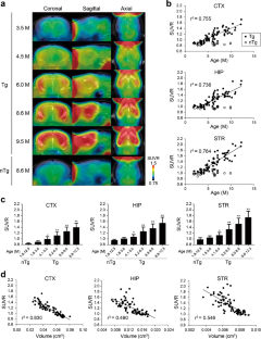
18F-PM-PBB3 SUVR at 40–60 min in the neocortex, hippocampus, and striatum of rTg4510 mice agreed with DVR, became significantly different from control values around 4–5 months of age, and progressively and negatively correlated with age and local volumes, respectively. Neocortical SUVR also correlated with the abundance of tau inclusions labeled with PM-PBB3 fluorescence, Gallyas-Braak silver impregnation, and anti-phospho-tau antibodies in postmortem assays. The in vivo and ex vivo 18F-PM-PBB3 binding was blocked by non-radioactive PM-PBB3. 18F-PM-PBB3 yielded a 1.6-fold greater dynamic range for tau imaging than its ancestor, 11C-PBB3.
Conclusion
Our imaging platform has enabled the quantification of tau depositions and consequent neuronal loss and is potentially applicable to the evaluation of candidate anti-tau and neuroprotective drugs.
中文翻译:
用于跟踪小鼠模型中病理性 tau 沉积和由此产生的神经元死亡的定量体内成像平台
目的
tau 原纤维的沉积与多种神经退行性疾病有关,包括阿尔茨海默病,对 tau 病理及其对神经元存活的影响的精确评估对于在有或没有潜在疗法的情况下研究神经退行性 tau 发病机制至关重要。我们旨在建立一个体内成像系统,通过正电子发射断层扫描 (PET) 量化 tau 积聚,并通过体积 MRI 量化 rTg4510 转基因小鼠模拟神经退行性 tau 病的脑萎缩。
方法
总共 91 只 rTg4510 和非转基因对照小鼠在不同年龄(1.8-12.3 个月)接受了带有 tau 放射性示踪剂、18 F-PM-PBB3 和 MRI 的 PET。使用小脑作为参考,目标区域中的放射性示踪剂结合被估计为标准化摄取值比(SUVR)和分布体积比(DVR)。还对来自扫描动物的脑切片进行组织病理学染色,以研究成像-神经病理学相关性。
结果
18 F-PM-PBB3 SUVR 在 40-60 分钟时在 rTg4510 小鼠的新皮层、海马体和纹状体中与 DVR 一致,在 4-5 个月大时与对照值显着不同,并且与年龄和局部逐渐呈负相关卷,分别。新皮质 SUVR 还与标有 PM-PBB3 荧光、Gallyas-Braak 银浸渍和死后检测中的抗磷酸化 tau 抗体的大量 tau 内含物相关。体内和体外18 F-PM-PBB3 结合被非放射性 PM-PBB3 阻断。18 F-PM-PBB3 产生的 tau 成像动态范围是其祖先11 C-PBB3的 1.6 倍。
结论
我们的成像平台能够量化 tau 沉积和随之而来的神经元丢失,并可能适用于候选抗 tau 药物和神经保护药物的评估。


 京公网安备 11010802027423号