当前位置:
X-MOL 学术
›
Microsc. Res. Tech.
›
论文详情
Our official English website, www.x-mol.net, welcomes your
feedback! (Note: you will need to create a separate account there.)
Liver in guinea pig: Scanning and transmission electron microscopic studies
Microscopy Research and Technique ( IF 2.0 ) Pub Date : 2022-07-04 , DOI: 10.1002/jemt.24194 Shunmugam Rajathi 1 , Geetha Ramesh 2 , Thandavan Arthanari Kannan 3 , Palanisamy Dharani 4 , Ravignanam Gnanadevi 5
Microscopy Research and Technique ( IF 2.0 ) Pub Date : 2022-07-04 , DOI: 10.1002/jemt.24194 Shunmugam Rajathi 1 , Geetha Ramesh 2 , Thandavan Arthanari Kannan 3 , Palanisamy Dharani 4 , Ravignanam Gnanadevi 5
Affiliation
|
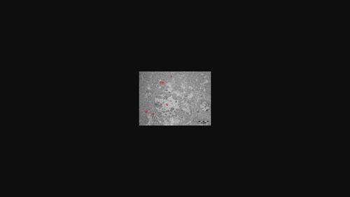
The liver performs many functions, which support metabolism, immunity, digestion, detoxification, vitamin storage, and so on. The liver is the storage organ for fat-soluble vitamins, iron and copper. Six adult healthy guinea pigs of 16–32 weeks of age (Irrespective of sex) were procured from the Department of Laboratory Animal Medicine, TANUVAS as per ethical committee approval. Animals were dissected as per CPCSEA norms and liver pieces were utilized for scanning electron microscopy (SEM) and transmission electron microscopy (TEM) study. The study was performed to document the ultrastructural details of liver of guinea pigs by SEM and TEM. By SEM, the liver parenchyma appeared as lobular with anastomosed one cell thick hepatic cell plates, which appeared as continuous sheets with small to large holes representing sinusoids. The sinusoidal lumen was lined by two types of cells namely endothelial cells and Kupffer cells. Endothelial cells had numerous large and small cytoplasmic fenestrations. Presence of Kupffer cells along with Ito cells were observed in the sinusoids. By TEM, hepatocytes had angular shape with sinusoidal face and bile canalicular face. Hepatocytes had oval nucleus with peripheral heterochromatin and central electron lucent granules and a prominent nucleolus. Bile canaliculi were formed by two adjacent hepatocytes. Fenestrated endothelial cells with flat and elongated shape were found to be in the formation of hepatic sinusoidal wall. A narrow space of Disse was found between the sinusoidal endothelium and hepatocytes. Kupffer cells and pit cells were found within the sinusoidal lumen. Ito cells were found in the space of Disse.
中文翻译:

豚鼠肝脏:扫描和透射电子显微镜研究
肝脏执行许多功能,支持新陈代谢、免疫、消化、解毒、维生素储存等。肝脏是脂溶性维生素、铁和铜的储存器官。根据伦理委员会的批准,从 TANUVAS 实验动物医学系采购了六只 16-32 周龄的成年健康豚鼠(不分性别)。根据 CPCSEA 规范解剖动物,并使用肝片进行扫描电子显微镜 (SEM) 和透射电子显微镜 (TEM) 研究。该研究旨在通过 SEM 和 TEM 记录豚鼠肝脏的超微结构细节。扫描电镜下,肝实质呈小叶状,吻合一细胞厚的肝细胞板,呈连续片状,由小到大的孔代表血窦。正弦腔内衬有两种类型的细胞,即内皮细胞和枯否细胞。内皮细胞有许多大大小小的细胞质开窗。在正弦曲线中观察到 Kupffer 细胞和 Ito 细胞的存在。透射电镜观察,肝细胞呈棱角状,呈正弦面和胆小管面。肝细胞核呈椭圆形,外周有异染色质,中央有电子透明颗粒和突出的核仁。胆小管由两个相邻的肝细胞形成。在肝窦壁的形成中发现了具有扁平和细长形状的有孔内皮细胞。在肝窦内皮和肝细胞之间发现狭窄的Disse间隙。在窦腔内发现了枯否细胞和凹坑细胞。在 Disse 空间中发现了 Ito 细胞。
更新日期:2022-07-04
中文翻译:
豚鼠肝脏:扫描和透射电子显微镜研究
肝脏执行许多功能,支持新陈代谢、免疫、消化、解毒、维生素储存等。肝脏是脂溶性维生素、铁和铜的储存器官。根据伦理委员会的批准,从 TANUVAS 实验动物医学系采购了六只 16-32 周龄的成年健康豚鼠(不分性别)。根据 CPCSEA 规范解剖动物,并使用肝片进行扫描电子显微镜 (SEM) 和透射电子显微镜 (TEM) 研究。该研究旨在通过 SEM 和 TEM 记录豚鼠肝脏的超微结构细节。扫描电镜下,肝实质呈小叶状,吻合一细胞厚的肝细胞板,呈连续片状,由小到大的孔代表血窦。正弦腔内衬有两种类型的细胞,即内皮细胞和枯否细胞。内皮细胞有许多大大小小的细胞质开窗。在正弦曲线中观察到 Kupffer 细胞和 Ito 细胞的存在。透射电镜观察,肝细胞呈棱角状,呈正弦面和胆小管面。肝细胞核呈椭圆形,外周有异染色质,中央有电子透明颗粒和突出的核仁。胆小管由两个相邻的肝细胞形成。在肝窦壁的形成中发现了具有扁平和细长形状的有孔内皮细胞。在肝窦内皮和肝细胞之间发现狭窄的Disse间隙。在窦腔内发现了枯否细胞和凹坑细胞。在 Disse 空间中发现了 Ito 细胞。


 京公网安备 11010802027423号