Nature Microbiology ( IF 20.5 ) Pub Date : 2022-06-30 , DOI: 10.1038/s41564-022-01164-2 David Chmielewski 1 , Michael F Schmid 2 , Graham Simmons 3, 4 , Jing Jin 3, 4 , Wah Chiu 1, 2, 5
|
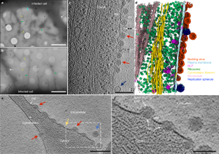
Chikungunya virus (CHIKV) is a representative alphavirus causing debilitating arthritogenic disease in humans. Alphavirus particles assemble into two icosahedral layers: the glycoprotein spike shell embedded in a lipid envelope and the inner nucleocapsid (NC) core. In contrast to matrix-driven assembly of some enveloped viruses, the assembly/budding process of two-layered icosahedral particles remains poorly understood. Here we used cryogenic electron tomography (cryo-ET) to capture snapshots of the CHIKV assembly in infected human cells. Subvolume classification of the snapshots revealed 12 intermediates representing different stages of assembly at the plasma membrane. Further subtomogram average structures ranging from subnanometre to nanometre resolutions show that immature non-icosahedral NCs function as rough scaffolds to trigger icosahedral assembly of the spike lattice, which in turn progressively transforms the underlying NCs into icosahedral cores during budding. Further, analysis of CHIKV-infected cells treated with budding-inhibiting antibodies revealed wider spaces between spikes than in icosahedral spike lattice, suggesting that spacing spikes apart to prevent their lateral interactions prevents the plasma membrane from bending around the NC, thus blocking virus budding. These findings provide the molecular mechanisms for alphavirus assembly and antibody-mediated budding inhibition that provide valuable insights for the development of broad therapeutics targeting the assembly of icosahedral enveloped viruses.
中文翻译:
使用低温电子断层扫描在原位可视化基孔肯雅病毒组装和出芽
基孔肯雅病毒 (CHIKV) 是一种代表性甲病毒,可导致人类衰弱性关节炎病。甲病毒颗粒组装成两个二十面体层:嵌入脂质包膜中的糖蛋白刺突壳和内部核衣壳 (NC) 核。与一些包膜病毒的矩阵驱动组装相比,双层二十面体颗粒的组装/出芽过程仍然知之甚少。在这里,我们使用低温电子断层扫描 (cryo-ET) 捕获受感染人类细胞中 CHIKV 组装的快照。快照的子体积分类揭示了 12 个中间体,代表质膜上组装的不同阶段。从亚纳米到纳米分辨率的进一步亚断层图平均结构表明,未成熟的非二十面体 NC 作为粗糙支架触发尖峰晶格的二十面体组装,这反过来在出芽过程中逐渐将底层 NC 转变为二十面体核心。此外,对用出芽抑制抗体处理的 CHIKV 感染细胞的分析显示,与二十面体尖峰晶格相比,尖峰之间的空间更宽,这表明间隔尖峰以防止它们的横向相互作用可防止质膜在 NC 周围弯曲,从而阻止病毒出芽。


 京公网安备 11010802027423号