当前位置:
X-MOL 学术
›
Microsc. Res. Tech.
›
论文详情
Our official English website, www.x-mol.net, welcomes your
feedback! (Note: you will need to create a separate account there.)
Automated bone healing evaluation: New approach to histomorphometric analysis
Microscopy Research and Technique ( IF 2.0 ) Pub Date : 2022-06-27 , DOI: 10.1002/jemt.24188 Camila Rodrigues Borges Linhares 1 , Gustavo Davi Rabelo 2 , Pedro Henrique Justino Oliveira Limirio 1 , Jessyca Figueira Venâncio 1 , Igor Gonçalves Ribeiro Silva 3 , Paula Dechichi 4
Microscopy Research and Technique ( IF 2.0 ) Pub Date : 2022-06-27 , DOI: 10.1002/jemt.24188 Camila Rodrigues Borges Linhares 1 , Gustavo Davi Rabelo 2 , Pedro Henrique Justino Oliveira Limirio 1 , Jessyca Figueira Venâncio 1 , Igor Gonçalves Ribeiro Silva 3 , Paula Dechichi 4
Affiliation
|
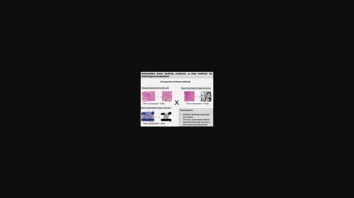
This study aimed to assess different approaches for bone healing evaluation on histological images and to introduce a new automatic evaluation method based on segmentation with distinct thresholds. We evaluated the hyperbaric oxygen therapy (HBO) effects on bone repair in type 1 diabetes mellitus rats. Twelve animals were divided into four groups (n = 3): non-diabetic, non-diabetic + HBO, diabetic, and diabetic + HBO. Diabetes was induced by intravenous administration of streptozotocin (50 mg/kg). Bone defects were created in femurs and HBO was immediately started at one session/day. After 7 days, the animals were euthanized, femurs were removed, demineralized, and embedded in paraffin. Histological sections were stained with hematoxylin and eosin (HE) and Mallory's trichrome (MT), and evaluated using three approaches: (1) conventional histomorphometric analysis (HE images) using a 144-point grid to quantify the bone matrix; (2) a semi-automatic method based on bone matrix segmentation to assess the bone matrix percentage (MT images); and (3) automatic approach, with the creation of a plug-in for ImageJ software. The time required to perform the analysis in each method was measured and subjected to Bland–Altman statistical analysis. All three methods were satisfactory for measuring bone formation and were not statistically different. The automatic approach reduced the working time compared to visual grid and semi-automated method (p < .01). Although histological evaluation of bone healing was performed successfully using all three methods, the novel automatic approach significantly shortened the time required for analysis and had high accuracy.
中文翻译:

自动骨愈合评估:组织形态计量分析的新方法
本研究旨在评估组织学图像上骨愈合评估的不同方法,并引入一种基于不同阈值分割的新自动评估方法。我们评估了高压氧治疗 (HBO) 对 1 型糖尿病大鼠骨修复的影响。十二只动物分为四组(n = 3):非糖尿病患者、非糖尿病患者 + HBO、糖尿病患者和糖尿病患者 + HBO。通过静脉注射链脲佐菌素 (50 mg/kg) 诱发糖尿病。在股骨中产生骨缺损,并立即开始 HBO,每天 1 次。7天后,将动物安乐死,取出股骨,脱矿质并包埋在石蜡中。组织切片用苏木精和伊红 (HE) 和马洛里三色 (MT) 染色,并使用三种方法进行评估:(1) 使用 144 点网格量化骨基质的常规组织形态计量分析(HE 图像);(2)一种基于骨基质分割的半自动评估骨基质百分比(MT图像)的方法;(3) 自动方法,为 ImageJ 软件创建插件。测量在每种方法中执行分析所需的时间并进行 Bland-Altman 统计分析。这三种方法对于测量骨形成都是令人满意的,并且没有统计学差异。与视觉网格和半自动方法相比,自动方法减少了工作时间(p < .01)。尽管使用所有三种方法都成功地进行了骨愈合的组织学评估,但新的自动方法显着缩短了分析所需的时间并具有很高的准确性。
更新日期:2022-06-27
中文翻译:
自动骨愈合评估:组织形态计量分析的新方法
本研究旨在评估组织学图像上骨愈合评估的不同方法,并引入一种基于不同阈值分割的新自动评估方法。我们评估了高压氧治疗 (HBO) 对 1 型糖尿病大鼠骨修复的影响。十二只动物分为四组(n = 3):非糖尿病患者、非糖尿病患者 + HBO、糖尿病患者和糖尿病患者 + HBO。通过静脉注射链脲佐菌素 (50 mg/kg) 诱发糖尿病。在股骨中产生骨缺损,并立即开始 HBO,每天 1 次。7天后,将动物安乐死,取出股骨,脱矿质并包埋在石蜡中。组织切片用苏木精和伊红 (HE) 和马洛里三色 (MT) 染色,并使用三种方法进行评估:(1) 使用 144 点网格量化骨基质的常规组织形态计量分析(HE 图像);(2)一种基于骨基质分割的半自动评估骨基质百分比(MT图像)的方法;(3) 自动方法,为 ImageJ 软件创建插件。测量在每种方法中执行分析所需的时间并进行 Bland-Altman 统计分析。这三种方法对于测量骨形成都是令人满意的,并且没有统计学差异。与视觉网格和半自动方法相比,自动方法减少了工作时间(p < .01)。尽管使用所有三种方法都成功地进行了骨愈合的组织学评估,但新的自动方法显着缩短了分析所需的时间并具有很高的准确性。


 京公网安备 11010802027423号