当前位置:
X-MOL 学术
›
Microsc. Res. Tech.
›
论文详情
Our official English website, www.x-mol.net, welcomes your
feedback! (Note: you will need to create a separate account there.)
Multi-scale attention network for segmentation of electron dense deposits in glomerular microscopic images
Microscopy Research and Technique ( IF 2.0 ) Pub Date : 2022-06-20 , DOI: 10.1002/jemt.24182 Jinyue Yang 1 , Xiuxiu Hu 2 , Hong Pan 3 , Pingsheng Chen 2 , Siyu Xia 1
Microscopy Research and Technique ( IF 2.0 ) Pub Date : 2022-06-20 , DOI: 10.1002/jemt.24182 Jinyue Yang 1 , Xiuxiu Hu 2 , Hong Pan 3 , Pingsheng Chen 2 , Siyu Xia 1
Affiliation
|
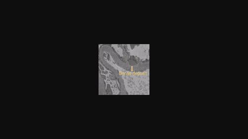
Electron dense deposit on the epithelial side of the glomerular capillary basement membrane is one of the pathological changes of membranous nephropathy. Automatic segmentation of deposits can relieve clinicians from the tedious and manual effort of identifying and localizing region of interest (ROI) in medical images and also assist to diagnose membranous nephropathy. Electron dense deposits are characterized by different sizes, irregular shapes, and low contrast to surrounding tissue structures in glomerular electron microscopy images. Considering the characteristics of dense deposits, we propose a multi-scale attention network for automatic segmentation of electron dense deposits of glomeruli in electron microscope images. Our method is built on the fully convolutional network but also takes advantages of the multi-scale skip connections and attention mechanism. Specifically, the multi-scale skip connection combines feature maps of different scales, makes the segmentation field larger, and integrates the shallow features of the image and high-level semantic information, which is more conducive to distinguishing dense deposits. At the same time, attention mechanism can focus on salient structures that normally produces a distinguishable feature representation. To evaluate the segmentation performance of the proposed method, we also collected a dataset of electron microscope images of membranous nephropathy. To the best of our knowledge, this is the largest image dataset for segmentation of glomerular basement membrane dense deposits. Experimental result shows that our model can accurately segment ordinary-sized dense deposits. Compared with state-of-the-art methods, our proposed method lower both false positive and false negative segmentation of small-sized protein sediments.
中文翻译:

用于分割肾小球显微图像中电子致密沉积物的多尺度注意力网络
肾小球毛细血管基底膜上皮侧电子致密沉积是膜性肾病的病理变化之一。沉积物的自动分割可以使临床医生从识别和定位医学图像中的感兴趣区域 (ROI) 的繁琐和手动工作中解脱出来,并且还有助于诊断膜性肾病。电子致密沉积物的特点是大小不一、形状不规则,在肾小球电子显微镜图像中与周围组织结构的对比度低。考虑到致密沉积物的特点,我们提出了一种多尺度注意力网络,用于自动分割电子显微镜图像中肾小球的电子致密沉积物。我们的方法建立在全卷积网络之上,但也利用了多尺度跳跃连接和注意机制。具体来说,多尺度skip connection结合了不同尺度的特征图,使得分割范围更大,同时融合了图像的浅层特征和高层语义信息,更有利于区分密集沉积物。同时,注意力机制可以专注于通常产生可区分特征表示的显着结构。为了评估所提出方法的分割性能,我们还收集了膜性肾病的电子显微镜图像数据集。据我们所知,这是用于分割肾小球基底膜致密沉积物的最大图像数据集。实验结果表明,我们的模型可以准确地分割普通大小的致密矿床。与最先进的方法相比,我们提出的方法降低了小尺寸蛋白质沉积物的假阳性和假阴性分割。
更新日期:2022-06-20
中文翻译:
用于分割肾小球显微图像中电子致密沉积物的多尺度注意力网络
肾小球毛细血管基底膜上皮侧电子致密沉积是膜性肾病的病理变化之一。沉积物的自动分割可以使临床医生从识别和定位医学图像中的感兴趣区域 (ROI) 的繁琐和手动工作中解脱出来,并且还有助于诊断膜性肾病。电子致密沉积物的特点是大小不一、形状不规则,在肾小球电子显微镜图像中与周围组织结构的对比度低。考虑到致密沉积物的特点,我们提出了一种多尺度注意力网络,用于自动分割电子显微镜图像中肾小球的电子致密沉积物。我们的方法建立在全卷积网络之上,但也利用了多尺度跳跃连接和注意机制。具体来说,多尺度skip connection结合了不同尺度的特征图,使得分割范围更大,同时融合了图像的浅层特征和高层语义信息,更有利于区分密集沉积物。同时,注意力机制可以专注于通常产生可区分特征表示的显着结构。为了评估所提出方法的分割性能,我们还收集了膜性肾病的电子显微镜图像数据集。据我们所知,这是用于分割肾小球基底膜致密沉积物的最大图像数据集。实验结果表明,我们的模型可以准确地分割普通大小的致密矿床。与最先进的方法相比,我们提出的方法降低了小尺寸蛋白质沉积物的假阳性和假阴性分割。


 京公网安备 11010802027423号