Our official English website, www.x-mol.net, welcomes your
feedback! (Note: you will need to create a separate account there.)
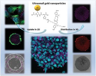
Uptake of Functional Ultrasmall Gold Nanoparticles in 3D Gut Cell Models
Small ( IF 13.0 ) Pub Date : 2022-06-16 , DOI: 10.1002/smll.202201167 Viktoriya Sokolova 1 , Jana-Fabienne Ebel 2 , Sebastian Kollenda 1 , Kai Klein 1 , Benedikt Kruse 1 , Claudia Veltkamp 3 , Christian M Lange 3 , Astrid M Westendorf 2 , Matthias Epple 1
Small ( IF 13.0 ) Pub Date : 2022-06-16 , DOI: 10.1002/smll.202201167 Viktoriya Sokolova 1 , Jana-Fabienne Ebel 2 , Sebastian Kollenda 1 , Kai Klein 1 , Benedikt Kruse 1 , Claudia Veltkamp 3 , Christian M Lange 3 , Astrid M Westendorf 2 , Matthias Epple 1
Affiliation
|
Ultrasmall gold nanoparticles (2 nm) easily penetrate the membranes of intestinal murine epithelial cells (MODE-K) and colorectal cancer cells (CT-26). They are also taken up by 3D spheroids (400 µm) of these cell types and primary gut organoids (500 µm). In contrast, dissolved dyes are not taken up by any of these cells or 3D structures. The distribution of fluorescent ultrasmall gold nanoparticles inside cells, spheroids, and gut organoids is examined by confocal laser scanning microscopy. Nanoparticles conjugated with the cytostatic drug doxorubicin and a fluorescent dye exhibit significantly greater cytotoxicity toward CT-26 tumor spheroids than equally concentrated dissolved doxorubicin, probably because they enter the interior of a spheroid much more easily than dissolved doxorubicin. Comprehensive analyses show that the cellular uptake of ultrasmall gold nanoparticles occurs by different endocytosis pathways.
中文翻译:

在 3D 肠道细胞模型中吸收功能性超小金纳米粒子
超小型金纳米粒子 (2 nm) 可轻松穿透肠鼠上皮细胞 (MODE-K) 和结肠直肠癌细胞 (CT-26) 的细胞膜。它们也被这些细胞类型的 3D 球体 (400 µm) 和主要肠道类器官 (500 µm) 吸收。相反,溶解的染料不会被任何这些细胞或 3D 结构吸收。通过共聚焦激光扫描显微镜检查荧光超小金纳米粒子在细胞、球体和肠道类器官内的分布。与细胞抑制药物多柔比星和荧光染料结合的纳米颗粒对 CT-26 肿瘤球体的细胞毒性显着高于同等浓度的溶解多柔比星,这可能是因为它们比溶解的多柔比星更容易进入球体内部。
更新日期:2022-06-16
中文翻译:
在 3D 肠道细胞模型中吸收功能性超小金纳米粒子
超小型金纳米粒子 (2 nm) 可轻松穿透肠鼠上皮细胞 (MODE-K) 和结肠直肠癌细胞 (CT-26) 的细胞膜。它们也被这些细胞类型的 3D 球体 (400 µm) 和主要肠道类器官 (500 µm) 吸收。相反,溶解的染料不会被任何这些细胞或 3D 结构吸收。通过共聚焦激光扫描显微镜检查荧光超小金纳米粒子在细胞、球体和肠道类器官内的分布。与细胞抑制药物多柔比星和荧光染料结合的纳米颗粒对 CT-26 肿瘤球体的细胞毒性显着高于同等浓度的溶解多柔比星,这可能是因为它们比溶解的多柔比星更容易进入球体内部。


 京公网安备 11010802027423号