Medical Image Analysis ( IF 10.7 ) Pub Date : 2022-06-05 , DOI: 10.1016/j.media.2022.102478 Ruobing Huang 1 , Mingrong Lin 1 , Haoran Dou 1 , Zehui Lin 1 , Qilong Ying 1 , Xiaohong Jia 2 , Wenwen Xu 2 , Zihan Mei 2 , Xin Yang 1 , Yijie Dong 2 , Jianqiao Zhou 2 , Dong Ni 1
|
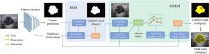
Breast Ultrasound (BUS) has proven to be an effective tool for the early detection of cancer in the breast. A lesion segmentation provides identification of the boundary, shape, and location of the target, and serves as a crucial step toward accurate diagnosis. Despite recent efforts in developing machine learning algorithms to automate this process, problems remain due to the blurry or occluded edges and highly irregular nodule shapes. Existing methods often produce over-smooth or inaccurate results, failing the need of identifying detailed boundary structures which are of clinical interest. To overcome these challenges, we propose a novel boundary-rendering framework that explicitly highlights the importance of boundary for automated nodule segmentation in BUS images. It utilizes a boundary selection module to automatically focuses on the ambiguous boundary region and a graph convolutional-based boundary rendering module to exploit global contour information. Furthermore, the proposed framework embeds nodule classification via semantic segmentation and encourages co-learning across tasks. Validation experiments were performed on different BUS datasets to verify the robustness of the proposed method. Results show that the proposed method outperforms states-of-art segmentation approaches (Dice=0.854, IOU=0.919, HD=17.8) in nodule delineation, as well as obtains a higher classification accuracy than classical classification models.
中文翻译:
用于超声图像中乳腺病变分割的边界渲染网络
乳房超声 (BUS) 已被证明是早期发现乳房癌症的有效工具。病灶分割可识别目标的边界、形状和位置,是准确诊断的关键步骤。尽管最近在开发机器学习算法以使该过程自动化,但由于边缘模糊或被遮挡以及高度不规则的结节形状,问题仍然存在。现有方法通常会产生过于平滑或不准确的结果,无法识别具有临床意义的详细边界结构。为了克服这些挑战,我们提出了一种新颖的边界渲染框架,该框架明确强调了边界对于 BUS 图像中自动结节分割的重要性。它利用边界选择模块自动关注模糊边界区域和基于图形卷积的边界渲染模块来利用全局轮廓信息。此外,所提出的框架通过语义分割嵌入结节分类,并鼓励跨任务的共同学习。在不同的 BUS 数据集上进行了验证实验,以验证所提出方法的鲁棒性。结果表明,所提出的方法在结节描绘方面优于最先进的分割方法(Dice=0.854,IOU=0.919,HD=17.8),并且获得了比经典分类模型更高的分类精度。提议的框架通过语义分割嵌入结节分类,并鼓励跨任务的共同学习。在不同的 BUS 数据集上进行了验证实验,以验证所提出方法的鲁棒性。结果表明,所提出的方法在结节描绘方面优于最先进的分割方法(Dice=0.854,IOU=0.919,HD=17.8),并且获得了比经典分类模型更高的分类精度。提议的框架通过语义分割嵌入结节分类,并鼓励跨任务的共同学习。在不同的 BUS 数据集上进行了验证实验,以验证所提出方法的鲁棒性。结果表明,所提出的方法在结节描绘方面优于最先进的分割方法(Dice=0.854,IOU=0.919,HD=17.8),并且获得了比经典分类模型更高的分类精度。


 京公网安备 11010802027423号