Drug Delivery and Translational Research ( IF 5.7 ) Pub Date : 2022-06-01 , DOI: 10.1007/s13346-022-01161-2 Parisa Ghandforoushan 1, 2 , Jalal Hanaee 2, 3 , Zahra Aghazadeh 4 , Mohammad Samiei 5 , Amir Mohammad Navali 6 , Ali Khatibi 7 , Soodabeh Davaran 1, 2, 8
|
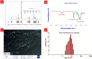
Since cartilage has a limited capacity for self-regeneration, treating cartilage degenerative disorders is a long-standing difficulty in orthopedic medicine. Researchers have scrutinized cartilage tissue regeneration to handle the deficiency of cartilage restoration capacity. This investigation proposed to compose an innovative nanocomposite biomaterial that enhances growth factor delivery to the injured cartilage site. Here, we describe the design and development of the biocompatible poly(lactide-co-glycolide) acid-collagen/poly(lactide-co-glycolide)-poly(ethylene glycol)-poly(lactide-co-glycolide) (PLGA-collagen/PLGA-PEG-PLGA) nanocomposite hydrogel containing transforming growth factor-β1 (TGF-β1). PLGA-PEG-PLGA nanoparticles were employed as a delivery system embedding TGF-β1 as an articular cartilage repair therapeutic agent. This study evaluates various physicochemical aspects of fabricated scaffolds by 1HNMR, FT-IR, SEM, BET, and DLS methods. The physicochemical features of the developed scaffolds, including porosity, density, degradation, swelling ratio, mechanical properties, morphologies, BET, ELISA, and cytotoxicity were assessed. The cell viability was investigated with the MTT test. Chondrogenic differentiation was assessed via Alcian blue staining and RT-PCR. In real-time PCR testing, the expression of Sox-9, collagen type II, and aggrecan genes was monitored. According to the results, human dental pulp stem cells (hDPSCs) exhibited high adhesion, proliferation, and differentiation on PLGA-collagen/PLGA-PEG-PLGA-TGFβ1 nanocomposite scaffolds compared to the control groups. SEM images displayed suitable cell adhesion and distribution of hDPSCs throughout the scaffolds. RT-PCR assay data displayed that TGF-β1 loaded PLGA-PEG-PLGA nanoparticles puts forward chondroblast differentiation in hDPSCs through the expression of chondrogenic genes. The findings revealed that PLGA-collagen/PLGA-PEG-PLGA-TGF-β1 nanocomposite hydrogel can be utilized as a supportive platform to support hDPSCs differentiation by implementing specific physio-chemical features.
Graphical abstract
中文翻译:
通过掺入载有 TGF-β1 的 PLGA-PEG-PLGA 纳米颗粒增强 PLGA 胶原支架的功能,用于使用人牙髓干细胞的软骨组织工程
由于软骨的自我再生能力有限,治疗软骨退行性疾病是骨科医学中长期存在的难题。研究人员仔细检查了软骨组织再生,以解决软骨修复能力不足的问题。这项研究提出了一种创新的纳米复合生物材料,可增强生长因子向受伤软骨部位的输送。在这里,我们描述了生物相容性聚(丙交酯-共-乙交酯)酸-胶原蛋白/聚(丙交酯-共-乙交酯)-聚(乙二醇)-聚(丙交酯-共-乙交酯)(PLGA-胶原蛋白)的设计和开发/PLGA-PEG-PLGA) 含有转化生长因子-β1 (TGF-β1) 的纳米复合水凝胶。PLGA-PEG-PLGA 纳米颗粒被用作嵌入 TGF-β1 作为关节软骨修复治疗剂的递送系统。1个HNMR、FT-IR、SEM、BET 和 DLS 方法。评估了所开发支架的物理化学特征,包括孔隙率、密度、降解、膨胀率、机械性能、形态、BET、ELISA 和细胞毒性。用MTT测试研究细胞活力。通过阿尔新蓝染色和 RT-PCR 评估软骨形成分化。在实时 PCR 测试中,监测了 Sox-9、II 型胶原蛋白和聚集蛋白聚糖基因的表达。根据结果,与对照组相比,人牙髓干细胞 (hDPSCs) 在 PLGA-胶原蛋白/PLGA-PEG-PLGA-TGFβ1 纳米复合支架上表现出高粘附、增殖和分化。SEM 图像显示 hDPSC 在整个支架中的适当细胞粘附和分布。RT-PCR 检测数据显示,载有 TGF-β1 的 PLGA-PEG-PLGA 纳米颗粒通过软骨形成基因的表达促进 hDPSC 中的成软骨细胞分化。研究结果表明,PLGA-胶原蛋白/PLGA-PEG-PLGA-TGF-β1 纳米复合水凝胶可用作支持平台,通过实现特定的物理化学特征来支持 hDPSCs 的分化。


 京公网安备 11010802027423号