Annals of Intensive Care ( IF 5.7 ) Pub Date : 2022-05-21 , DOI: 10.1186/s13613-022-01013-9 Jean Pasqueron 1 , Pauline Dureau 1 , Gauthier Arcile 1 , Baptiste Duceau 1 , Geoffroy Hariri 1 , Victoria Lepère 1 , Guillaume Lebreton 2 , Jean-Jacques Rouby 3 , Adrien Bouglé 1
|
Background
Hospital-acquired pneumonia (HAP) is the most common and severe complication in patients treated with venoarterial extracorporeal membrane oxygenation (VA ECMO) and its diagnosis remains challenging. Nothing is known about the usefulness of lung ultrasound (LUS) in early detection of HAP in patients treated with VA ECMO. Also, LUS and chest radiography were performed when HAP was suspected in cardiac critically ill adult VA ECMO presenting with acute respiratory failure. The sonographic features of HAP in VA ECMO patients were determined and we assessed the performance of the lung ultrasound simplified clinical pulmonary score (LUS-sCPIS), the sCPIS and bioclinical parameters or chest radiography alone for early diagnosis of HAP.
Results
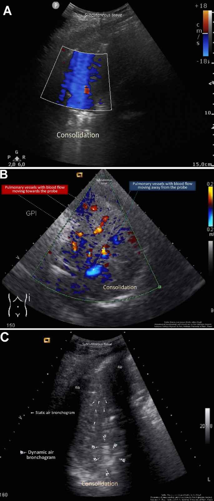
We included 70 patients, of which 44 (63%) were independently diagnosed with HAP. LUS examination revealed that color Doppler intrapulmonary flow (P = 0.0000043) and dynamic air bronchogram (P = 0.00024) were the most frequent HAP-related signs. The LUS-sCPIS (area under the curve = 0.77) yielded significantly better results than the sCPIS (area under the curve = 0.65; P = 0.004), while leukocyte count, temperature and chest radiography were not discriminating for HAP diagnosis.
Discussion
Diagnosis of HAP is a daily challenge for the clinician managing patients on venoarterial ECMO. Lung ultrasound can be a valuable tool as the initial imaging modality for the diagnosis of pneumonia. Color Doppler intrapulmonary flow and dynamic air bronchogram appear to be particularly insightful for the diagnosis of HAP.
中文翻译:
肺超声在早期发现静脉动脉体外膜肺氧合心脏重症患者医院获得性肺炎中的应用
背景
医院获得性肺炎(HAP)是接受静脉动脉体外膜肺氧合(VA ECMO)治疗的患者最常见和最严重的并发症,其诊断仍然具有挑战性。对于接受 VA ECMO 治疗的患者,肺部超声 (LUS) 在早期发现 HAP 中的作用尚不清楚。此外,在出现急性呼吸衰竭的心脏危重成人 VA ECMO 中怀疑 HAP 时,还进行了 LUS 和胸片检查。确定了 VA ECMO 患者中 HAP 的超声特征,我们评估了肺部超声简化临床肺部评分 (LUS-sCPIS)、sCPIS 和生物临床参数或胸部 X 光片的表现,以早期诊断 HAP。
结果
我们纳入了 70 名患者,其中 44 名(63%)独立诊断为 HAP。LUS 检查显示,彩色多普勒肺内血流 ( P = 0.0000043 ) 和动态空气支气管图 ( P = 0.00024 ) 是最常见的 HAP 相关体征。LUS-sCPIS(曲线下面积 = 0.77)比 sCPIS(曲线下面积 = 0.65;P = 0.004)产生显着更好的结果,而白细胞计数、温度和胸部 X 光片不能区分 HAP 诊断。
讨论
HAP 的诊断是临床医生管理静脉动脉 ECMO 患者的日常挑战。肺部超声可以作为一种有价值的工具,作为诊断肺炎的初始成像方式。彩色多普勒肺内血流和动态空气支气管图似乎对 HAP 的诊断特别有洞察力。


 京公网安备 11010802027423号