当前位置:
X-MOL 学术
›
Microsc. Res. Tech.
›
论文详情
Our official English website, www.x-mol.net, welcomes your
feedback! (Note: you will need to create a separate account there.)
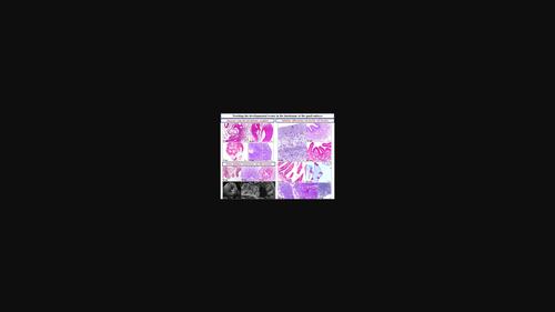
Tracking the developmental events in the duodenum of the quail embryo: Using light and electron microscope
Microscopy Research and Technique ( IF 2.0 ) Pub Date : 2022-05-12 , DOI: 10.1002/jemt.24146 Fatma Abdelhakeem 1, 2 , Salma A Mohamed 2 , Mohammed Abdelsabour-Khalaf 2 , Fatma A Madkour 2
Microscopy Research and Technique ( IF 2.0 ) Pub Date : 2022-05-12 , DOI: 10.1002/jemt.24146 Fatma Abdelhakeem 1, 2 , Salma A Mohamed 2 , Mohammed Abdelsabour-Khalaf 2 , Fatma A Madkour 2
Affiliation
|
The present study described the full morphology of the duodenum of the Japanese quail during the embryonic stage from 3rd day of incubation till hatching using the light and electron (scanning and transmission) microscope. The specimens were collected, analyzed and described anatomically, morphometrically and microscopically. The first recognition of the prospective duodenum was at the 4th day of incubation and developed continuously by age progression. The prospective duodenum consisted of a flat pseudostratified epithelium, mesenchyme and covering mesothelium. On day 8th of incubation, the epithelium developed three evaginations lead to formation three previllous ridges protruding inside the duodenal lumen, which later at the 9th day differentiated into numbers of projections; villi. On the 9th day, the epithelium lined the villi transformed into a simple columnar type, the duodenal villi appeared as pyramidal-shaped projections, had wide base and narrow apex and by age progression, the duodenal villi went through changes in number, size and shape. On hatching day, the duodenal epithelium consisted of enterocytes interspersed with secretory goblet cells, which stained positive for both Periodic Acid Schiff (PAS) and Alcian blue AB and represented filled with metachromatic granules. The muscular wall started as mesenchymal condensation on the 6th day then differentiated into the circular smooth muscle layer on the 9th day of incubation. Giving detailed information about the morphological development of the duodenum during the incubation period of quail embryo helps in reaching a satisfactory explanation about how the duodenum plays a vital role in digestion, absorption and immunity.
中文翻译:

跟踪鹌鹑胚胎十二指肠的发育事件:使用光学和电子显微镜
本研究使用光学和电子(扫描和透射)显微镜描述了日本鹌鹑从孵化第 3 天到孵化期间十二指肠的完整形态。收集、分析和描述解剖学、形态学和显微镜下的标本。预期十二指肠的第一次识别是在孵化的第 4 天,并随着年龄的增长而不断发展。预期的十二指肠由扁平的假复层上皮、间质和覆盖间皮组成。孵化第8天,上皮出现3个外翻,形成3个突出于十二指肠腔内的绒毛前嵴,第9天后分化为多个突起;绒毛 第 9 天,衬在绒毛上的上皮转变为简单的柱状,十二指肠绒毛呈锥体状突起,基部宽,顶端窄,随着年龄的增长,十二指肠绒毛的数量、大小和形状发生变化。孵化当天,十二指肠上皮由肠细胞组成,散布着分泌杯状细胞,高碘酸席夫(PAS)和阿尔新蓝AB染色呈阳性,并充满异染颗粒。肌肉壁在第 6 天开始为间充质浓缩,然后在孵化第 9 天分化为圆形平滑肌层。
更新日期:2022-05-12
中文翻译:
跟踪鹌鹑胚胎十二指肠的发育事件:使用光学和电子显微镜
本研究使用光学和电子(扫描和透射)显微镜描述了日本鹌鹑从孵化第 3 天到孵化期间十二指肠的完整形态。收集、分析和描述解剖学、形态学和显微镜下的标本。预期十二指肠的第一次识别是在孵化的第 4 天,并随着年龄的增长而不断发展。预期的十二指肠由扁平的假复层上皮、间质和覆盖间皮组成。孵化第8天,上皮出现3个外翻,形成3个突出于十二指肠腔内的绒毛前嵴,第9天后分化为多个突起;绒毛 第 9 天,衬在绒毛上的上皮转变为简单的柱状,十二指肠绒毛呈锥体状突起,基部宽,顶端窄,随着年龄的增长,十二指肠绒毛的数量、大小和形状发生变化。孵化当天,十二指肠上皮由肠细胞组成,散布着分泌杯状细胞,高碘酸席夫(PAS)和阿尔新蓝AB染色呈阳性,并充满异染颗粒。肌肉壁在第 6 天开始为间充质浓缩,然后在孵化第 9 天分化为圆形平滑肌层。


 京公网安备 11010802027423号