Our official English website, www.x-mol.net, welcomes your
feedback! (Note: you will need to create a separate account there.)
High-throughput digital pathology via a handheld, multiplexed, and AI-powered ptychographic whole slide scanner
Lab on a Chip ( IF 6.1 ) Pub Date : 2022-05-12 , DOI: 10.1039/d2lc00084a Shaowei Jiang 1 , Chengfei Guo 1 , Pengming Song 1 , Tianbo Wang 1 , Ruihai Wang 1 , Terrance Zhang 1 , Qian Wu 2 , Rishikesh Pandey 1 , Guoan Zheng 1
Lab on a Chip ( IF 6.1 ) Pub Date : 2022-05-12 , DOI: 10.1039/d2lc00084a Shaowei Jiang 1 , Chengfei Guo 1 , Pengming Song 1 , Tianbo Wang 1 , Ruihai Wang 1 , Terrance Zhang 1 , Qian Wu 2 , Rishikesh Pandey 1 , Guoan Zheng 1
Affiliation
|
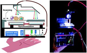
The recent advent of whole slide imaging (WSI) systems has moved digital pathology closer to diagnostic applications and clinical practices. Integrating WSI with machine learning promises the growth of this field in upcoming years. Here we report the design and implementation of a handheld, colour-multiplexed, and AI-powered ptychographic whole slide scanner for digital pathology applications. This handheld scanner is built using low-cost and off-the-shelf components, including red, green, and blue laser diodes for sample illumination, a modified stage for programmable sample positioning, and a synchronized image sensor pair for data acquisition. We smear a monolayer of goat blood cells on the main sensor for high-resolution lensless coded ptychographic imaging. The synchronized secondary sensor acts as a non-contact encoder for precisely tracking the absolute object position for ptychographic reconstruction. For WSI, we introduce a new phase-contrast-based focus metric for post-acquisition autofocusing of both stained and unstained specimens. We show that the scanner can resolve the 388-nm linewidth on the resolution target and acquire gigapixel images with a 14 mm × 11 mm area in ∼70 seconds. The imaging performance is validated with regular stained pathology slides, unstained thyroid smears, and malaria-infected blood smears. The deep neural network developed in this study further enables high-throughput cytometric analysis using the recovered complex amplitude. The reported do-it-yourself scanner offers a portable solution to transform the high-end WSI system into one that can be made widely available at a low cost. The capability of high-throughput quantitative phase imaging may also find applications in rapid on-site evaluations.
中文翻译:

通过手持式、多路复用和人工智能驱动的 ptychographic 全玻片扫描仪实现高通量数字病理学
最近出现的全玻片成像 (WSI) 系统使数字病理学更接近诊断应用和临床实践。将 WSI 与机器学习相结合,有望在未来几年推动该领域的发展。在这里,我们报告了用于数字病理学应用的手持式、彩色多路复用和人工智能驱动的 ptychographic 全玻片扫描仪的设计和实施。这种手持式扫描仪使用低成本和现成的组件构建,包括用于样品照明的红色、绿色和蓝色激光二极管、用于可编程样品定位的改进阶段以及用于数据采集的同步图像传感器对。我们在主传感器上涂抹单层山羊血细胞,以进行高分辨率无透镜编码 ptychographic 成像。同步辅助传感器充当非接触式编码器,用于精确跟踪绝对对象位置以进行 ptychographic 重建。对于 WSI,我们引入了一种新的基于相位对比的对焦指标,用于染色和未染色标本的采集后自动对焦。我们表明,扫描仪可以在分辨率目标上解析 388 nm 线宽,并在 70 秒内获取 14 mm × 11 mm 区域的千兆像素图像。成像性能通过常规染色病理切片、未染色甲状腺涂片和感染疟疾的血液涂片进行验证。本研究中开发的深度神经网络进一步实现了使用恢复的复振幅进行高通量细胞计数分析。报告的自己动手扫描仪提供了一种便携式解决方案,可将高端 WSI 系统转变为可以以低成本广泛使用的系统。高通量定量相位成像的能力也可以在快速现场评估中找到应用。
更新日期:2022-05-12
中文翻译:
通过手持式、多路复用和人工智能驱动的 ptychographic 全玻片扫描仪实现高通量数字病理学
最近出现的全玻片成像 (WSI) 系统使数字病理学更接近诊断应用和临床实践。将 WSI 与机器学习相结合,有望在未来几年推动该领域的发展。在这里,我们报告了用于数字病理学应用的手持式、彩色多路复用和人工智能驱动的 ptychographic 全玻片扫描仪的设计和实施。这种手持式扫描仪使用低成本和现成的组件构建,包括用于样品照明的红色、绿色和蓝色激光二极管、用于可编程样品定位的改进阶段以及用于数据采集的同步图像传感器对。我们在主传感器上涂抹单层山羊血细胞,以进行高分辨率无透镜编码 ptychographic 成像。同步辅助传感器充当非接触式编码器,用于精确跟踪绝对对象位置以进行 ptychographic 重建。对于 WSI,我们引入了一种新的基于相位对比的对焦指标,用于染色和未染色标本的采集后自动对焦。我们表明,扫描仪可以在分辨率目标上解析 388 nm 线宽,并在 70 秒内获取 14 mm × 11 mm 区域的千兆像素图像。成像性能通过常规染色病理切片、未染色甲状腺涂片和感染疟疾的血液涂片进行验证。本研究中开发的深度神经网络进一步实现了使用恢复的复振幅进行高通量细胞计数分析。报告的自己动手扫描仪提供了一种便携式解决方案,可将高端 WSI 系统转变为可以以低成本广泛使用的系统。高通量定量相位成像的能力也可以在快速现场评估中找到应用。


 京公网安备 11010802027423号