JACC: Cardiovascular Imaging ( IF 12.8 ) Pub Date : 2022-05-11 , DOI: 10.1016/j.jcmg.2022.02.029 Giovanni Peretto 1 , Elena Busnardo 2 , Paola Ferro 3 , Anna Palmisano 4 , Davide Vignale 4 , Antonio Esposito 4 , Giacomo De Luca 5 , Corrado Campochiaro 6 , Silvia Sartorelli 6 , Monica De Gaspari 7 , Stefania Rizzo 7 , Lorenzo Dagna 8 , Cristina Basso 7 , Luigi Gianolli 3 , Paolo Della Bella 9 , Simone Sala 10
|
Background
18F-fluorodeoxyglucose positron emission tomography (FDG-PET) scan has no recognized role in diagnosis, prognosis, and disease monitoring in patients with arrhythmic myocarditis.
Objectives
The purpose of this study was to investigate the value of FDG-PET scan in arrhythmic myocarditis.
Methods
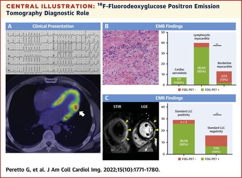
The authors enrolled 75 consecutive patients (age 47 ± 14 years, 65% men) undergoing FDG-PET scan for arrhythmic myocarditis. Myocarditis was diagnosed by endomyocardial biopsy (EMB) and, whenever applicable, cardiac magnetic resonance (CMR).
Results
Indications for FDG-PET scan included either contraindication to CMR (n = 50) or mismatch between CMR and EMB (n = 25). Overall, 50 patients (67%) had positive FDG-PET. Sensitivity was 75% referring to EMB, and 73% to CMR. Specificity was 67% referring to EMB, and 59% to CMR. FDG-PET accuracy was lower in the presence of borderline myocarditis, and either late (>30 days) or on-immunosuppression FDG-PET scanning. Anteroseptal distribution pattern, found in 12 of 50 (24%) patients including 7 of 7 cardiac sarcoidosis cases, was associated with greater occurrence of ventricular arrhythmias and atrioventricular blocks in 4.2 ± 1.7 years of follow-up (10 of 12 vs 7 of 38, and 7 of 12 vs 0 of 38, respectively; both P < 0.001). In 39 patients (52%), FDG-PET was repeated by 13 ± 2 months, allowing immunosuppression withdrawal after FDG uptake normalization either by first (76%) or second reassessment (24%).
Conclusions
FDG-PET scan may be a clinically useful diagnostic technique in arrhythmic myocarditis, in particular when CMR is unsuitable because of irregular heartbeat or implantable cardioverter-defibrillator–related artifacts. Anteroseptal FDG distribution is associated with a worse arrhythmic outcome and should raise the suspicion of cardiac sarcoidosis. During follow-up, repeated FDG-PET allows myocarditis monitoring to guide immunosuppression withdrawal.
中文翻译:
FDG-PET扫描在心律失常性心肌炎中的临床应用
背景
18 F-氟脱氧葡萄糖正电子发射断层扫描 (FDG-PET) 扫描在心律失常性心肌炎患者的诊断、预后和疾病监测中没有公认的作用。
目标
本研究的目的是探讨 FDG-PET 扫描在心律失常性心肌炎中的价值。
方法
作者连续招募了 75 名接受 FDG-PET 扫描的心律失常性心肌炎患者(年龄 47 ± 14 岁,65% 为男性)。心肌炎通过心内膜心肌活检 (EMB) 诊断,并且在适用的情况下,通过心脏磁共振 (CMR) 诊断。
结果
FDG-PET 扫描的适应症包括 CMR 的禁忌症 (n = 50) 或 CMR 和 EMB 之间的不匹配 (n = 25)。总体而言,50 名患者 (67%) 的 FDG-PET 呈阳性。对 EMB 的敏感性为 75%,对 CMR 的敏感性为 73%。EMB 的特异性为 67%,CMR 的特异性为 59%。FDG-PET 准确性在存在临界性心肌炎、晚期(>30 天)或免疫抑制 FDG-PET 扫描时较低。在 4.2 ± 1.7 年的随访中,50 名患者中有 12 名(24%)发现了前间隔分布模式,包括 7 名心脏结节病病例中的 7 名,这与室性心律失常和房室传导阻滞的发生率较高相关(12 人中有 10 人,38 人中有 7 人) , 和 7 of 12 vs 0 of 38, 分别; 两者P <0.001)。在 39 名患者 (52%) 中,FDG-PET 重复 13 ± 2 个月,允许在 FDG 摄取正常化后通过第一次 (76%) 或第二次重新评估 (24%) 停用免疫抑制。
结论
FDG-PET 扫描可能是临床上对心律失常性心肌炎有用的诊断技术,特别是当由于心律不齐或植入式心脏复律除颤器相关伪影而导致 CMR 不适用时。前间隔 FDG 分布与较差的心律失常结果相关,应引起对心脏结节病的怀疑。在随访期间,重复的 FDG-PET 允许监测心肌炎以指导免疫抑制的退出。


 京公网安备 11010802027423号