Cell Biology and Toxicology ( IF 5.3 ) Pub Date : 2022-05-04 , DOI: 10.1007/s10565-022-09712-6 Wanda van der Stel 1 , Huan Yang 1 , Sylvia E le Dévédec 1 , Bob van de Water 1 , Joost B Beltman 1 , Erik H J Danen 1
|
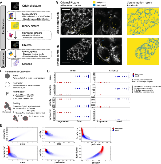
Cells can adjust their mitochondrial morphology by altering the balance between mitochondrial fission and fusion to adapt to stressful conditions. The connection between a chemical perturbation, changes in mitochondrial function, and altered mitochondrial morphology is not well understood. Here, we made use of high-throughput high-content confocal microscopy to assess the effects of distinct classes of oxidative phosphorylation (OXPHOS) complex inhibitors on mitochondrial parameters in a concentration and time resolved manner. Mitochondrial morphology phenotypes were clustered based on machine learning algorithms and mitochondrial integrity patterns were mapped. In parallel, changes in mitochondrial membrane potential (MMP), mitochondrial and cellular ATP levels, and viability were microscopically assessed. We found that inhibition of MMP, mitochondrial ATP production, and oxygen consumption rate (OCR) using sublethal concentrations of complex I and III inhibitors did not trigger mitochondrial fragmentation. Instead, complex V inhibitors that suppressed ATP and OCR but increased MMP provoked a more fragmented mitochondrial morphology. In agreement, complex V but not complex I or III inhibitors triggered proteolytic cleavage of the mitochondrial fusion protein, OPA1. The relation between increased MMP and fragmentation did not extend beyond OXPHOS complex inhibitors: increasing MMP by blocking the mPTP pore did not lead to OPA1 cleavage or mitochondrial fragmentation and the OXPHOS uncoupler FCCP was associated with OPA1 cleavage and MMP reduction. Altogether, our findings connect vital mitochondrial functions and phenotypes in a high-throughput high-content confocal microscopy approach that help understanding of chemical-induced toxicity caused by OXPHOS complex perturbing chemicals.
中文翻译:
高内涵高通量成像揭示了 OXPHOS 复合物 I、III 和 V 抑制剂的线粒体形态和功能之间的明显联系
细胞可以通过改变线粒体裂变和融合之间的平衡来调整线粒体形态,以适应压力条件。化学扰动、线粒体功能变化和线粒体形态改变之间的联系尚不清楚。在这里,我们利用高通量高内涵共聚焦显微镜以浓度和时间分辨的方式评估不同类别的氧化磷酸化 (OXPHOS) 复合物抑制剂对线粒体参数的影响。基于机器学习算法对线粒体形态表型进行聚类,并绘制了线粒体完整性模式图。同时,线粒体膜电位 (MMP)、线粒体和细胞 ATP 水平以及活力的变化在显微镜下进行了评估。我们发现抑制 MMP,使用亚致死浓度的复合物 I 和 III 抑制剂的线粒体 ATP 产生和耗氧率 (OCR) 不会引发线粒体断裂。相反,抑制 ATP 和 OCR 但增加 MMP 的复合物 V 抑制剂会引发更加支离破碎的线粒体形态。同意,复合物 V 而不是复合物 I 或 III 抑制剂触发线粒体融合蛋白 OPA1 的蛋白水解切割。MMP 增加与碎片化之间的关系并未超出 OXPHOS 复合物抑制剂:通过阻断 mPTP 孔增加 MMP 不会导致 OPA1 裂解或线粒体碎片化,OXPHOS 解偶联剂 FCCP 与 OPA1 裂解和 MMP 减少有关。共,


 京公网安备 11010802027423号