Cell Biology and Toxicology ( IF 5.3 ) Pub Date : 2022-04-29 , DOI: 10.1007/s10565-022-09714-4 Jing Ma 1 , Yi Li 1 , Mengxuan Chen 1 , Weihang Wang 1 , Qiqian Zhao 1 , Bo He 1 , Min Zhang 1 , Yongfang Jiang 1
|
Objective
To investigate the effects of human bone marrow mesenchymal stem cells (hMSCs)-derived exosome circCDK13 on liver fibrosis and its mechanism.
Methods
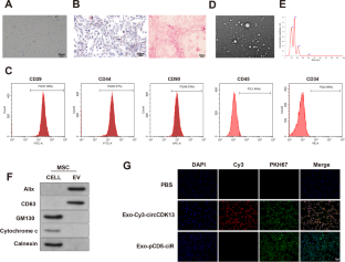
Exosomes derived from hMSCs were extracted and identified by flow cytometry and osteogenic and adipogenic induction, and the expressions of marker proteins on the surface of exosomes were detected by western blot. Cell proliferation was measured by CCK8 assay, the expression of active markers of HSCs by immunofluorescence, and the expressions of fibrosis-related factors by western blot. A mouse model of liver fibrosis was established by intraperitoneal injection of thioacetamide (TAA). Fibrosis was detected by HE staining, Masson staining, and Sirius red staining. Western blot was utilized to test the expressions of PI3K/AKT and NF-κB pathway related proteins, dual-luciferase reporter assay and RIP assay to validate the binding between circCDK13 and miR-17-5p as well as between miR-17-5p and KAT2B, and ChIP to validate the effect of KAT2B on H3 acetylation and MFGE8 transcription.
Results
hMSCs-derived exosomes inhibited liver fibrosis mainly through circCDK13. Dual-luciferase reporter assay and RIP assay demonstrated the binding between circCDK13 and miR-17-5p as well as between miR-17-5p and KAT2B. Further experimental results indicated that circCDK13 mediated liver fibrosis by regulating the miR-17-5p/KAT2B axis, and KAT2B promoted MFGE8 transcription by H3 acetylation. Exo-circCDK13 inhibited PI3K/AKT and NF-κB signaling pathways activation through regulating the miR-17-5p/KAT2B axis.
Conclusion
hMSCs-derived exosome circCDK13 inhibited liver fibrosis by regulating the expression of MFGE8 through miR-17-5p/KAT2B axis.
Graphical abstract
中文翻译:
hMSCs来源的外泌体circCDK13通过miR-17-5p/KAT2B调控MFGE8表达抑制肝纤维化
客观的
探讨人骨髓间充质干细胞(hMSCs)来源的外泌体circCDK13对肝纤维化的影响及其机制。
方法
提取源自hMSCs的外泌体,通过流式细胞术和成骨和脂肪诱导进行鉴定,并通过western blot检测外泌体表面标记蛋白的表达。CCK8检测细胞增殖,免疫荧光检测HSCs活性标志物的表达,western blot检测纤维化相关因子的表达。通过腹腔注射硫代乙酰胺(TAA)建立小鼠肝纤维化模型。通过HE染色、Masson染色和天狼星红染色检测纤维化。Western blot检测PI3K/AKT和NF-κB通路相关蛋白的表达,双荧光素酶报告实验和RIP实验验证circCDK13与miR-17-5p以及miR-17-5p与miR-17-5p的结合。卡特2B,
结果
hMSCs来源的外泌体主要通过circCDK13抑制肝纤维化。双荧光素酶报告分析和 RIP 分析证明了 circCDK13 和 miR-17-5p 之间以及 miR-17-5p 和 KAT2B 之间的结合。进一步的实验结果表明,circCDK13通过调控miR-17-5p/KAT2B轴介导肝纤维化,KAT2B通过H3乙酰化促进MFGE8转录。Exo-circCDK13 通过调节 miR-17-5p/KAT2B 轴抑制 PI3K/AKT 和 NF-κB 信号通路的激活。
结论
hMSCs 衍生的外泌体 circCDK13 通过 miR-17-5p/KAT2B 轴调节 MFGE8 的表达来抑制肝纤维化。


 京公网安备 11010802027423号