Immunity ( IF 25.5 ) Pub Date : 2022-04-20 , DOI: 10.1016/j.immuni.2022.03.020 Sizun Jiang 1 , Chi Ngai Chan 2 , Xavier Rovira-Clavé 3 , Han Chen 3 , Yunhao Bai 3 , Bokai Zhu 3 , Erin McCaffrey 3 , Noah F Greenwald 3 , Candace Liu 3 , Graham L Barlow 3 , Jason L Weirather 4 , John Paul Oliveria 5 , Tsuguhisa Nakayama 6 , Ivan T Lee 7 , Matthias S Matter 8 , Anne E Carlisle 4 , Darci Philips 3 , Gustavo Vazquez 3 , Nilanjan Mukherjee 3 , Kathleen Busman-Sahay 2 , Michael Nekorchuk 2 , Margaret Terry 2 , Skyler Younger 2 , Marc Bosse 3 , Janos Demeter 3 , Scott J Rodig 9 , Alexandar Tzankov 8 , Yury Goltsev 3 , David Robert McIlwain 3 , Michael Angelo 3 , Jacob D Estes 10 , Garry P Nolan 3
|
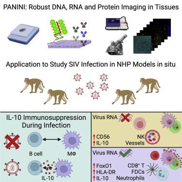
Understanding the mechanisms of HIV tissue persistence necessitates the ability to visualize tissue microenvironments where infected cells reside; however, technological barriers limit our ability to dissect the cellular components of these HIV reservoirs. Here, we developed protein and nucleic acid in situ imaging (PANINI) to simultaneously quantify DNA, RNA, and protein levels within these tissue compartments. By coupling PANINI with multiplexed ion beam imaging (MIBI), we measured over 30 parameters simultaneously across archival lymphoid tissues from healthy or simian immunodeficiency virus (SIV)-infected nonhuman primates. PANINI enabled the spatial dissection of cellular phenotypes, functional markers, and viral events resulting from infection. SIV infection induced IL-10 expression in lymphoid B cells, which correlated with local macrophage M2 polarization. This highlights a potential viral mechanism for conditioning an immunosuppressive tissue environment for virion production. The spatial multimodal framework here can be extended to decipher tissue responses in other infectious diseases and tumor biology.
中文翻译:
蛋白质和核酸联合成像揭示了组织微环境中病毒依赖性 B 细胞和巨噬细胞的免疫抑制
了解 HIV 组织持续存在的机制需要能够可视化受感染细胞所在的组织微环境;然而,技术障碍限制了我们剖析这些艾滋病毒储存库细胞成分的能力。在这里,我们开发了蛋白质和核酸原位成像 (PANINI),以同时量化这些组织区室中的 DNA、RNA 和蛋白质水平。通过将 PANINI 与多重离子束成像 (MIBI) 结合,我们同时测量了健康或感染猿猴免疫缺陷病毒 (SIV) 的非人灵长类动物的档案淋巴组织的 30 多个参数。 PANINI 能够对细胞表型、功能标记和感染引起的病毒事件进行空间剖析。 SIV 感染诱导淋巴 B 细胞中 IL-10 的表达,这与局部巨噬细胞 M2 极化相关。这凸显了调节免疫抑制组织环境以产生病毒粒子的潜在病毒机制。这里的空间多模态框架可以扩展到破译其他传染病和肿瘤生物学中的组织反应。


 京公网安备 11010802027423号