Cell Stem Cell ( IF 19.8 ) Pub Date : 2022-03-10 , DOI: 10.1016/j.stem.2022.02.008 Stephen Gadomski 1 , Claire Fielding 2 , Andrés García-García 2 , Claudia Korn 2 , Chrysa Kapeni 2 , Sadaf Ashraf 3 , Javier Villadiego 4 , Raquel Del Toro 5 , Olivia Domingues 6 , Jeremy N Skepper 7 , Tatiana Michel 6 , Jacques Zimmer 6 , Regine Sendtner 8 , Scott Dillon 9 , Kenneth E S Poole 10 , Gill Holdsworth 11 , Michael Sendtner 8 , Juan J Toledo-Aral 4 , Cosimo De Bari 3 , Andrew W McCaskie 12 , Pamela G Robey 13 , Simón Méndez-Ferrer 14
|
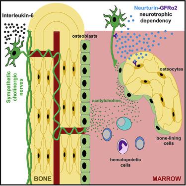
The autonomic nervous system is a master regulator of homeostatic processes and stress responses. Sympathetic noradrenergic nerve fibers decrease bone mass, but the role of cholinergic signaling in bone has remained largely unknown. Here, we describe that early postnatally, a subset of sympathetic nerve fibers undergoes an interleukin-6 (IL-6)-induced cholinergic switch upon contacting the bone. A neurotrophic dependency mediated through GDNF-family receptor-α2 (GFRα2) and its ligand, neurturin (NRTN), is established between sympathetic cholinergic fibers and bone-embedded osteocytes, which require cholinergic innervation for their survival and connectivity. Bone-lining osteoprogenitors amplify and propagate cholinergic signals in the bone marrow (BM). Moderate exercise augments trabecular bone partly through an IL-6-dependent expansion of sympathetic cholinergic nerve fibers. Consequently, loss of cholinergic skeletal innervation reduces osteocyte survival and function, causing osteopenia and impaired skeletal adaptation to moderate exercise. These results uncover a cholinergic neuro-osteocyte interface that regulates skeletogenesis and skeletal turnover through bone-anabolic effects.
中文翻译:
胆碱能神经骨骼界面促进出生后生长和运动期间的骨形成
自主神经系统是体内平衡过程和应激反应的主要调节器。交感去甲肾上腺素能神经纤维会减少骨量,但胆碱能信号在骨中的作用仍然很大程度上未知。在这里,我们描述了出生后早期,一部分交感神经纤维在接触骨骼时经历白细胞介素 6 (IL-6) 诱导的胆碱能转换。交感胆碱能纤维和骨嵌入骨细胞之间建立了通过 GDNF 家族受体 α2 (GFRα2) 及其配体神经营养因子 (NRTN) 介导的神经营养依赖性,骨细胞的生存和连接需要胆碱能神经支配。骨内骨祖细胞放大并传播骨髓 (BM) 中的胆碱能信号。适度运动部分通过 IL-6 依赖性交感胆碱能神经纤维扩张来增强骨小梁。因此,胆碱能骨骼神经支配的丧失会降低骨细胞的存活和功能,导致骨质减少和骨骼对适度运动的适应受损。这些结果揭示了胆碱能神经骨细胞界面,通过骨合成代谢作用调节骨骼发生和骨骼更新。


 京公网安备 11010802027423号