Clinical Spectroscopy Pub Date : 2022-02-18 , DOI: 10.1016/j.clispe.2022.100021 Callum Gassner 1 , John A. Adegoke 1 , Sheila K. Patel 2 , Varun J. Sharma 3, 4 , Kamila Kochan 1 , Louise M. Burrell 2 , Jaishankar Raman 3, 4 , Bayden R. Wood 1
|
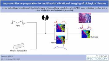
The complementary nature of Infrared (IR) and Raman spectroscopies enables a thorough understanding of biological tissue – so called multimodal vibrational spectroscopic imaging. However, new approaches in terms of sample preparation and data analysis are required to release the full potential of multimodal spectroscopy. Herein, we propose an inexpensive and relatively simple sample preparation technique incorporating mirror-finished stainless-steel slides and polyethylene glycol as an embedding medium that is compatible for both infrared and Raman spectroscopy of tissue sections. K-Means Clustering and Principal Component Analysis (PCA) were used to evaluate the performance of multimodal vibrational spectroscopic imaging compared with IR and Raman spectroscopic imaging individually using a rat kidney as a model. The K-Means cluster maps generated with the multimodal dataset showed the best correlation between different tissue types identified by an adjacent section stained with Masson’s Trichrome compared to either Raman or IR spectroscopy analysed independently. PCA score maps of the multimodal dataset produced a clear separation of individual tissue types along the first three Principal Components. Additionally, PCA permitted the correlation of IR and Raman peaks arising mainly from collagen vibrational modes. Finally, polyethylene glycol embedding is shown as an attractive alternative to paraffin embedding for spectroscopic analyses, due to significantly less fluorescence in Raman measurements and retention of lipids in the tissue, without any retention of the medium within the tissue.
中文翻译:
用于生物组织多模态振动成像的改进组织制备
红外 (IR) 和拉曼光谱的互补性使我们能够彻底了解生物组织——即所谓的多模态振动光谱成像。然而,需要样品制备和数据分析方面的新方法来释放多模态光谱学的全部潜力。在这里,我们提出了一种廉价且相对简单的样品制备技术,将镜面不锈钢载玻片和聚乙二醇作为嵌入介质,与组织切片的红外和拉曼光谱兼容。K-Means 聚类和主成分分析 (PCA) 用于评估多模态振动光谱成像与单独使用大鼠肾脏作为模型的 IR 和拉曼光谱成像的性能。与独立分析的拉曼或红外光谱相比,使用多模式数据集生成的 K-Means 聚类图显示了由用马森三色染色的相邻切片识别的不同组织类型之间的最佳相关性。多模式数据集的 PCA 得分图沿前三个主成分产生了单个组织类型的清晰分离。此外,PCA 允许主要由胶原振动模式产生的 IR 和拉曼峰的相关性。最后,聚乙二醇包埋被证明是一种有吸引力的替代石蜡包埋的光谱分析方法,因为拉曼测量中的荧光显着减少,并且组织中的脂质保留,而组织内没有任何介质保留。


 京公网安备 11010802027423号