JACC: Cardiovascular Imaging ( IF 12.8 ) Pub Date : 2021-12-15 , DOI: 10.1016/j.jcmg.2021.09.029 Vasken Dilsizian 1 , Ricardo P J Budde 2 , Wengen Chen 1 , Sunil V Mankad 3 , Jonathan R Lindner 4 , Koen Nieman 5
|
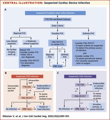
The diagnosis of cardiac device infection and, more importantly, accurate localization of the infection site, such as defibrillator pocket, pacemaker lead, along the peripheral driveline or central portion of the left ventricular assist device, prosthetic valve ring abscesses, and perivalvular extensions, remain clinically challenging. Although transthoracic and transesophageal echocardiography are the first-line imaging tests in suspected endocarditis and for assessing hemodynamic complications, recent studies suggest that cardiac computed tomography (CT) or CT angiography and functional imaging with 18F-fluoro-2-deoxyglucose (FDG) positron emission tomography (PET) with CT (FDG PET/CT) may have an incremental role in technically limited or inconclusive cases on echocardiography. One of the key benefits of FDG PET/CT is in its detection of inflammatory cells early in the infection process, before morphological damages ensue. However, there are many unanswered questions in the literature. In this document, we provide consensus on best practices among the various imaging studies, which includes the detection of cardiac device infection, differentiation of infection from inflammation, image-guided patient management, and detailed recommendations on patient preparation, image acquisition, processing, interpretation, and standardized reporting.
中文翻译:
成像心脏装置相关感染和心内膜炎的最佳实践
心脏装置感染的诊断,更重要的是,感染部位的准确定位,如除颤器口袋、起搏器导线、左心室辅助装置的外围传动系统或中心部分、人工瓣环脓肿和瓣周延伸,仍然存在临床具有挑战性。虽然经胸和经食道超声心动图是疑似心内膜炎和评估血流动力学并发症的一线影像学检查,但最近的研究表明,心脏计算机断层扫描 (CT) 或 CT 血管造影和功能成像18F-氟-2-脱氧葡萄糖 (FDG) 正电子发射断层扫描 (PET) 与 CT (FDG PET/CT) 可能在超声心动图技术受限或不确定的病例中发挥重要作用。FDG PET/CT 的主要优势之一是在感染过程的早期检测炎症细胞,在形态损伤发生之前。然而,文献中有许多悬而未决的问题。在本文档中,我们就各种影像学研究的最佳实践达成共识,其中包括心脏装置感染的检测、感染与炎症的鉴别、影像引导的患者管理以及对患者准备、图像采集、处理、解释的详细建议和标准化的报告。


 京公网安备 11010802027423号