International Journal of Cardiology ( IF 3.2 ) Pub Date : 2021-11-20 , DOI: 10.1016/j.ijcard.2021.11.048 Patrick Langguth 1 , Mona Salehi Ravesh 1 , Jörg Detlev Moritz 1 , Katy Rinne 2 , Paul Lennard Harneit 2 , Joshua Kian Khodami 2 , Joachim Graessner 3 , Anselm Uebing 4 , Olav Jansen 1 , Marcus Both 1 , Jan Hinnerk Hansen 4
|
Objectives
To evaluate the ability of non-contrast enhanced magnetic resonance imaging (MRI) techniques to characterize Fontan associated liver disease (FALD) in adolescent and adult Fontan patients.
Methods
Fontan patients (n = 29) and healthy controls (n = 13) underwent an MRI protocol with T1, T2 and Apparent Diffusion Coefficient (ADC) mapping. Routine FALD screening included abdominal ultrasound and laboratory testing.
Results
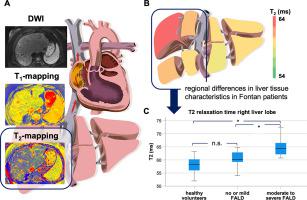
Median follow-up after Fontan operation was 15.1 (IQR 12.0–16.8) years. Distinct differences in tissue characteristics were visualized. T1 and T2 relaxation times were prolonged in Fontan patients, particularly of the right lobe (T1: 745 (IQR 715–784) ms vs. 586 (IQR 555–602) ms, p < 0.001; T2: 63 (IQR 59–64) ms vs. 58 (IQR 56–60) ms, p = 0.002). Left lobe ADC was lower in Fontan patients (1.10 (IQR 1.06–1.18) x 10−3 mm2/s vs. 1.23 (IQR 1.19–1.29) x 10−3 mm2/s, p < 0.001). T2 mapping was able to differentiate between controls and Fontan patients with different FALD severity. Right lobe T2 was higher in patients with moderate or severe in comparison to those with no or mild changes and healthy controls (64 (IQR 61–67) ms vs. 60 (IQR 59–63) ms vs. 58 (IQR 56–60) ms, p = 0.001).
Conclusions
Non-contrast enhanced MRI methods are able to visualize regional differences in liver tissue characteristics. T1 and T2 relaxation times were prolonged in Fontan patients suggestive of fibrosis or congestive hepatopathy, while reduced ADC might reflect impaired microperfusion. These methods have promising clinical potential for detection of liver abnormalities in Fontan patients. The usefulness of T2 mapping to grade FALD severity merits further investigation.
中文翻译:
用于表征 Fontan 相关肝病的非对比增强磁共振成像
目标
评估非对比增强磁共振成像 (MRI) 技术在青少年和成人 Fontan 患者中表征 Fontan 相关肝病 (FALD) 的能力。
方法
Fontan 患者 (n = 29) 和健康对照 (n = 13) 接受了具有 T 1、 T 2和表观扩散系数 (ADC) 映射的 MRI 方案。常规 FALD 筛查包括腹部超声和实验室检测。
结果
Fontan 手术后的中位随访时间为 15.1 (IQR 12.0–16.8) 年。可见组织特征的明显差异。Fontan 患者的T 1和 T 2弛豫时间延长,尤其是右叶 (T 1 : 745 (IQR 715–784) ms vs. 586 (IQR 555–602) ms, p < 0.001; T 2 : 63 ( IQR 59–64) 毫秒与 58 (IQR 56–60) 毫秒,p = 0.002)。Fontan 患者的左叶 ADC 较低 (1.10 (IQR 1.06–1.18) x 10 -3 mm 2 /s vs. 1.23 (IQR 1.19–1.29) x 10 -3 mm 2 /s, p < 0.001)。T 2映射能够区分具有不同 FALD 严重程度的对照组和 Fontan 患者。右叶 T 2 中度或重度患者与没有或轻度变化的患者和健康对照组相比更高(64(IQR 61-67)ms vs. 60(IQR 59-63)ms vs. 58(IQR 56-60)ms, p = 0.001)。
结论
非对比增强 MRI 方法能够可视化肝组织特征的区域差异。提示纤维化或充血性肝病的 Fontan 患者的T 1和 T 2弛豫时间延长,而 ADC 降低可能反映微灌注受损。这些方法在检测 Fontan 患者的肝脏异常方面具有广阔的临床潜力。T 2映射到 FALD 严重程度的有用性值得进一步研究。


 京公网安备 11010802027423号