当前位置:
X-MOL 学术
›
ACS Photonics
›
论文详情
Our official English website, www.x-mol.net, welcomes your
feedback! (Note: you will need to create a separate account there.)
Multiplane Encoded Light-Sheet Microscopy for Enhanced 3D Imaging
ACS Photonics ( IF 6.5 ) Pub Date : 2021-11-03 , DOI: 10.1021/acsphotonics.1c01401 Alessandro Zunino 1, 2 , Francesco Garzella 1, 3 , Alberta Trianni 1, 2 , Peter Saggau 1, 4 , Paolo Bianchini 1 , Alberto Diaspro 1, 2 , Martí Duocastella 1, 5
ACS Photonics ( IF 6.5 ) Pub Date : 2021-11-03 , DOI: 10.1021/acsphotonics.1c01401 Alessandro Zunino 1, 2 , Francesco Garzella 1, 3 , Alberta Trianni 1, 2 , Peter Saggau 1, 4 , Paolo Bianchini 1 , Alberto Diaspro 1, 2 , Martí Duocastella 1, 5
Affiliation
|
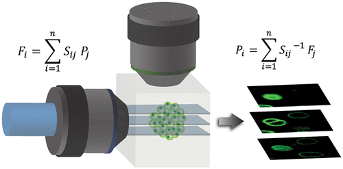
Light-sheet microscopes have become the tool of choice for volumetric imaging of large samples. Based on a wide-field acquisition scheme, they are capable of optical sectioning at diffraction-limited resolution and minimal overall photodamage. Unfortunately, traditional architectures are limited in speed because 3D images are collected by either sample translation or synchronized movement of both light-sheet and detection objective lens. A promising solution avoiding slow mechanical movements is to extend the depth-of-field of the microscope and moving only the light-sheet. However, this normally comes at the cost of losing light and contrast, compromising the signal-to-noise ratio of the images. Here, we propose an innovative technique devoted to restoring the quality of the images, while preserving the speed of extended depth-of-field microscopes. It is based on generating a stack of parallel light-sheets using a pair of orthogonal acousto-optic deflectors, enabling the simultaneous illumination of different sample planes. Given the extended depth-of-field, all such planes appear in focus and can be acquired in a superimposed single frame. By applying a single-step inversion algorithm, we can decode a stack of frames into a volumetric image whose signal-to-noise ratio and contrast are greatly enhanced. We provide a detailed theoretical framework of the method and demonstrate its feasibility with volumetric images of kidney cell spheroids.
中文翻译:

用于增强 3D 成像的多平面编码光片显微镜
光片显微镜已成为大型样品体积成像的首选工具。基于宽视场采集方案,它们能够以衍射极限分辨率和最小的整体光损伤进行光学切片。不幸的是,传统架构的速度受到限制,因为 3D 图像是通过样本平移或光片和检测物镜的同步运动来收集的。避免缓慢机械运动的一个有前途的解决方案是扩展显微镜的景深并仅移动光片。然而,这通常以损失光和对比度为代价,从而影响图像的信噪比。在这里,我们提出了一种致力于恢复图像质量的创新技术,同时保持扩展景深显微镜的速度。它基于使用一对正交声光偏转器生成一叠平行光片,从而能够同时照明不同的样品平面。鉴于扩展的景深,所有这些平面都出现在焦点上,并且可以在叠加的单帧中获取。通过应用单步反演算法,我们可以将一堆帧解码成一个体积图像,其信噪比和对比度大大增强。我们提供了该方法的详细理论框架,并通过肾细胞球体的体积图像证明了其可行性。鉴于扩展的景深,所有这些平面都出现在焦点上,并且可以在叠加的单帧中获取。通过应用单步反演算法,我们可以将一堆帧解码成一个体积图像,其信噪比和对比度大大增强。我们提供了该方法的详细理论框架,并通过肾细胞球体的体积图像证明了其可行性。鉴于扩展的景深,所有这些平面都出现在焦点上,并且可以在叠加的单帧中获取。通过应用单步反演算法,我们可以将一堆帧解码成一个体积图像,其信噪比和对比度大大增强。我们提供了该方法的详细理论框架,并通过肾细胞球体的体积图像证明了其可行性。
更新日期:2021-11-17
中文翻译:
用于增强 3D 成像的多平面编码光片显微镜
光片显微镜已成为大型样品体积成像的首选工具。基于宽视场采集方案,它们能够以衍射极限分辨率和最小的整体光损伤进行光学切片。不幸的是,传统架构的速度受到限制,因为 3D 图像是通过样本平移或光片和检测物镜的同步运动来收集的。避免缓慢机械运动的一个有前途的解决方案是扩展显微镜的景深并仅移动光片。然而,这通常以损失光和对比度为代价,从而影响图像的信噪比。在这里,我们提出了一种致力于恢复图像质量的创新技术,同时保持扩展景深显微镜的速度。它基于使用一对正交声光偏转器生成一叠平行光片,从而能够同时照明不同的样品平面。鉴于扩展的景深,所有这些平面都出现在焦点上,并且可以在叠加的单帧中获取。通过应用单步反演算法,我们可以将一堆帧解码成一个体积图像,其信噪比和对比度大大增强。我们提供了该方法的详细理论框架,并通过肾细胞球体的体积图像证明了其可行性。鉴于扩展的景深,所有这些平面都出现在焦点上,并且可以在叠加的单帧中获取。通过应用单步反演算法,我们可以将一堆帧解码成一个体积图像,其信噪比和对比度大大增强。我们提供了该方法的详细理论框架,并通过肾细胞球体的体积图像证明了其可行性。鉴于扩展的景深,所有这些平面都出现在焦点上,并且可以在叠加的单帧中获取。通过应用单步反演算法,我们可以将一堆帧解码成一个体积图像,其信噪比和对比度大大增强。我们提供了该方法的详细理论框架,并通过肾细胞球体的体积图像证明了其可行性。


 京公网安备 11010802027423号