Seminars in Thoracic and Cardiovascular Surgery ( IF 2.6 ) Pub Date : 2021-10-22 , DOI: 10.1053/j.semtcvs.2021.10.007 Bilguun Erkhem-Ochir 1 , Wataru Tatsuishi 2 , Takehiko Yokobori 3 , Navchaa Gombodorj 4 , Hiroshi Saeki 5 , Ken Shirabe 5 , Tomonobu Abe 2
|
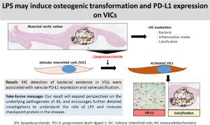
Aortic stenosis (AS) is a disease characterized by narrowing of the aortic valve (AV) orifice. The purpose of this study was to clarify the significance of bacterial detection and clinicopathological factors, including valve-infiltrating immune cells and disease severity, in relation to AS. After obtaining the written informed consent form from 50 patients with AS, we performed immunohistochemical analysis of lipopolysaccharide (LPS) for gram-negative bacteria and lipoteichoic acid (LTA) for gram-positive bacteria on surgically resected-AVs. Moreover, we evaluated the relationships among the presence of bacteria, immune checkpoint protein PD-L1 expression, and immune cell infiltrations such as CD8-positive T lymphocytes, CD163-positive macrophages, and FOXP3-positive regulatory T cell (Treg) in resected-aortic valves. LPS detection in the resected-aortic valve tissues was significantly associated with stromal PD-L1 expression, valve calcification, and LTA existence in resected samples. We showed that the presence of LPS was significantly related to high PD-L1 expression only in calcified-AVs, not in non-calcified-AVs. Moreover, the high expression of PD-L1 in AS samples without LPS was significantly associated with positive infiltration of CD163-positive macrophages and FOXP3-positive Tregs. Immunohistochemical bacterial detection in resected-aortic valves was associated with PD-L1 accumulation and valve calcification. PD-L1 significantly accumulated only in calcified valves with LPS existence.
中文翻译:
切除瓣膜中细菌的免疫组织化学检测与可能导致钙化性主动脉瓣狭窄的基质免疫检查点蛋白表达相关
主动脉瓣狭窄 (AS) 是一种以主动脉瓣(AV) 口变窄为特征的疾病。本研究的目的是阐明与 AS 相关的细菌检测和临床病理因素(包括瓣膜浸润免疫细胞和疾病严重程度)的重要性。在获得50 名 AS 患者的书面知情同意书后,我们对手术切除的 AV 上的革兰氏阴性菌脂多糖 (LPS) 和革兰氏阳性菌脂磷壁酸 (LTA) 进行了免疫组织化学分析。此外,我们评估了细菌的存在、免疫检查点蛋白 PD-L1 表达和免疫细胞浸润(如 CD8 阳性)之间的关系切除主动脉瓣中的 T 淋巴细胞、CD163 阳性巨噬细胞和 FOXP3 阳性调节性 T 细胞 (Treg)。切除的主动脉瓣组织中的 LPS 检测与基质 PD-L1 表达、瓣膜钙化和切除样本中 LTA 的存在显着相关。我们表明,LPS 的存在仅与钙化 AV 中的高 PD-L1 表达显着相关,而非钙化 AV 中。此外,不含 LPS 的 AS 样本中 PD-L1 的高表达与 CD163 阳性巨噬细胞和 FOXP3 阳性 Treg 的阳性浸润显着相关。切除的主动脉瓣中的免疫组织化学细菌检测与 PD-L1 积聚和瓣膜钙化有关。PD-L1 仅在存在 LPS 的钙化瓣膜中显着积累。


 京公网安备 11010802027423号