Medical Image Analysis ( IF 10.7 ) Pub Date : 2021-10-04 , DOI: 10.1016/j.media.2021.102230 Duyan Ta 1 , Yanshuai Tu 1 , Zhong-Lin Lu 2 , Yalin Wang 1
|
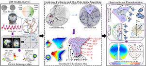
The retinotopic map depicts the cortical neurons’ response to visual stimuli on the retina and has contributed significantly to our understanding of human visual system. Although recent advances in high field functional magnetic resonance imaging (fMRI) have made it possible to generate the in vivo retinotopic map with great detail, quantifying the map remains challenging. Existing quantification methods do not preserve surface topology and often introduce large geometric distortions to the map. In this study, we developed a new framework based on computational conformal geometry and quasiconformal Teichmüller theory to quantify the retinotopic map. Specifically, we introduced a general pipeline, consisting of cortical surface conformal parameterization, surface-spline-based cortical activation signal smoothing, and vertex-wise Beltrami coefficient-based map description. After correcting most of the violations of the topological conditions, the result was a “Beltrami coefficient map” (BCM) that rigorously and completely characterizes the retinotopic map by quantifying the local quasiconformal mapping distortion at each visual field location. The BCM provided topological and fully reconstructable retinotopic maps. We successfully applied the new framework to analyze the V1 retinotopic maps from the Human Connectome Project (n=181), the largest state of the art retinotopy dataset currently available. With unprecedented precision, we found that the V1 retinotopic map was quasiconformal and the local mapping distortions were similar across observers. The new framework can be applied to other visual areas and retinotopic maps of individuals with and without eye diseases, and improve our understanding of visual cortical organization in normal and clinical populations.
中文翻译:
基于准共形映射的人类视网膜专题图的定量表征
视网膜专题图描绘了皮质神经元对视网膜上视觉刺激的反应,对我们对人类视觉系统的理解做出了重大贡献。尽管高场功能磁共振成像(fMRI)的最新进展使得生成非常详细的体内视网膜专题图成为可能,但量化该图仍然具有挑战性。现有的量化方法不能保留表面拓扑,并且经常会给地图带来较大的几何扭曲。在这项研究中,我们开发了一个基于计算共形几何和准共形 Teichmüller 理论的新框架来量化视网膜专题图。具体来说,我们引入了一个通用流程,包括皮质表面共形参数化、基于表面样条的皮质激活信号平滑和基于顶点的贝尔特拉米系数的映射描述。在纠正了大部分违反拓扑条件的情况后,结果是“贝尔特拉米系数图”(BCM),它通过量化每个视野位置的局部准共形映射失真来严格、完整地表征视网膜专题图。BCM 提供拓扑和完全可重建的视网膜专题图。我们成功应用新框架来分析来自人类连接组计划 (n=181) 的 V1 视网膜专题图,这是目前可用的最先进的视网膜专题数据集。以前所未有的精度,我们发现 V1 视网膜专题图是准共形的,并且观察者之间的局部映射失真是相似的。新框架可应用于患有和未患有眼病的个体的其他视觉区域和视网膜专题图,并提高我们对正常和临床人群视觉皮层组织的理解。


 京公网安备 11010802027423号