当前位置:
X-MOL 学术
›
Biomater. Sci.
›
论文详情
Our official English website, www.x-mol.net, welcomes your
feedback! (Note: you will need to create a separate account there.)
The in vivo fate of tobacco mosaic virus nanoparticle theranostic agents modified by the addition of a polydopamine coat
Biomaterials Science ( IF 5.8 ) Pub Date : 2021-09-30 , DOI: 10.1039/d1bm01113h Christian Isalomboto Nkanga 1 , Young Hun Chung 2 , Sourabh Shukla 1 , Jingcheng Zhou 1 , Jesse V Jokerst 1, 3, 4 , Nicole F Steinmetz 1, 2, 4, 5, 6, 7
Biomaterials Science ( IF 5.8 ) Pub Date : 2021-09-30 , DOI: 10.1039/d1bm01113h Christian Isalomboto Nkanga 1 , Young Hun Chung 2 , Sourabh Shukla 1 , Jingcheng Zhou 1 , Jesse V Jokerst 1, 3, 4 , Nicole F Steinmetz 1, 2, 4, 5, 6, 7
Affiliation
|
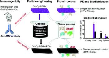
Plant virus nanoparticles (VNPs) have multiple advantages over their synthetic counterparts including the cost-effective large-scale manufacturing of uniform particles that are easy to functionalize. Tobacco mosaic virus (TMV) is one of the most promising VNP scaffolds, reflecting its high aspect ratio and ability to carry and/or display multivalent therapeutic ligands and contrast agents. Here we investigated the circulation, protein corona, immunogenicity, and organ distribution/clearance of TMV particles internally co-labeled with cyanine 5 (Cy5) and chelated gadolinium (Gd) for dual tracking by fluorescence imaging and optical emission spectrometry, with or without an external coating of polydopamine (PDA) to confer photothermal and photoacoustic capabilities. The PDA-coated particles (Gd-Cy5-TMV-PDA) showed a shorter plasma circulation time and broader distribution to organs of the reticuloendothelial system (liver, lungs, and spleen) than uncoated Gd-Cy5-TMV particles (liver and spleen only). The Gd-Cy5-TMV-PDA particles were surrounded by 2–10-fold greater protein corona (containing mainly immunoglobulins) compared to Gd-Cy5-TMV particles. However, the enzyme-linked immunosorbent assay (ELISA) revealed that PDA-coated particles bind 2-fold lesser to anti-TMV antibodies elicited by particle injection than uncoated particles, suggesting that the PDA coat enables evasion from systemic antibody surveillance. Gd-Cy5-TMV-PDA particles were cleared from organs after 8 days compared to 5 days for the uncoated particles. The slower tissue clearance of the coated particles makes them ideal for theranostic applications by facilitating sustained local delivery in addition to multimodal imaging and photothermal capabilities. We have demonstrated the potential of PDA-coated proteinaceous nanoparticles for multiple biomedical applications.
中文翻译:

通过添加聚多巴胺涂层修饰的烟草花叶病毒纳米颗粒治疗诊断剂的体内命运
植物病毒纳米颗粒(VNP)比其合成同类产品具有多种优势,包括经济高效地大规模制造易于功能化的均匀颗粒。烟草花叶病毒 (TMV) 是最有前途的 VNP 支架之一,反映了其高纵横比以及携带和/或展示多价治疗配体和造影剂的能力。在这里,我们研究了内部与花青 5 (Cy5) 和螯合钆 (Gd) 共标记的 TMV 颗粒的循环、蛋白冠、免疫原性和器官分布/清除,通过荧光成像和光学发射光谱法进行双重跟踪,无论有或没有聚多巴胺 (PDA) 外部涂层可赋予光热和光声功能。与未包被的 Gd-Cy5-TMV 颗粒(仅肝脏和脾脏)相比,PDA 包被的颗粒(Gd-Cy5-TMV-PDA)表现出更短的血浆循环时间和更广泛的网状内皮系统器官(肝、肺和脾)分布。 )。与 Gd-Cy5-TMV 颗粒相比,Gd-Cy5-TMV-PDA 颗粒被 2-10 倍大的蛋白冠(主要含有免疫球蛋白)包围。然而,酶联免疫吸附测定 (ELISA) 显示,PDA 包被的颗粒与颗粒注射引起的抗 TMV 抗体的结合比未包被的颗粒少 2 倍,这表明 PDA 包被能够逃避全身抗体监测。 Gd-Cy5-TMV-PDA 颗粒在 8 天后从器官中清除,而未包被的颗粒则需要 5 天。涂层颗粒的组织清除速度较慢,除了多模态成像和光热功能外,还促进持续的局部递送,使其成为治疗诊断应用的理想选择。 我们已经证明了 PDA 包被的蛋白质纳米颗粒在多种生物医学应用中的潜力。
更新日期:2021-09-30
中文翻译:
通过添加聚多巴胺涂层修饰的烟草花叶病毒纳米颗粒治疗诊断剂的体内命运
植物病毒纳米颗粒(VNP)比其合成同类产品具有多种优势,包括经济高效地大规模制造易于功能化的均匀颗粒。烟草花叶病毒 (TMV) 是最有前途的 VNP 支架之一,反映了其高纵横比以及携带和/或展示多价治疗配体和造影剂的能力。在这里,我们研究了内部与花青 5 (Cy5) 和螯合钆 (Gd) 共标记的 TMV 颗粒的循环、蛋白冠、免疫原性和器官分布/清除,通过荧光成像和光学发射光谱法进行双重跟踪,无论有或没有聚多巴胺 (PDA) 外部涂层可赋予光热和光声功能。与未包被的 Gd-Cy5-TMV 颗粒(仅肝脏和脾脏)相比,PDA 包被的颗粒(Gd-Cy5-TMV-PDA)表现出更短的血浆循环时间和更广泛的网状内皮系统器官(肝、肺和脾)分布。 )。与 Gd-Cy5-TMV 颗粒相比,Gd-Cy5-TMV-PDA 颗粒被 2-10 倍大的蛋白冠(主要含有免疫球蛋白)包围。然而,酶联免疫吸附测定 (ELISA) 显示,PDA 包被的颗粒与颗粒注射引起的抗 TMV 抗体的结合比未包被的颗粒少 2 倍,这表明 PDA 包被能够逃避全身抗体监测。 Gd-Cy5-TMV-PDA 颗粒在 8 天后从器官中清除,而未包被的颗粒则需要 5 天。涂层颗粒的组织清除速度较慢,除了多模态成像和光热功能外,还促进持续的局部递送,使其成为治疗诊断应用的理想选择。 我们已经证明了 PDA 包被的蛋白质纳米颗粒在多种生物医学应用中的潜力。


 京公网安备 11010802027423号