Our official English website, www.x-mol.net, welcomes your feedback! (Note: you will need to create a separate account there.)
Improvement of biomolecular analysis in thin films using in situ matrix enhanced secondary ion mass spectrometry
Analyst ( IF 4.2 ) Pub Date : 2021-08-30 , DOI: 10.1039/d1an00727k Konstantin Moshkunov 1 , Benjamin Tomasetti 1 , Thomas Daphnis 1 , Vincent Delmez 1 , Kevin Vanvarenberg 2 , Véronique Préat 2 , Matthias Lorenz 3, 4 , Jusal Quanico 5 , Geert Baggerman 5, 6 , Filip Lemiere 5, 7 , Christine Dupont 1 , Arnaud Delcorte 1
Analyst ( IF 4.2 ) Pub Date : 2021-08-30 , DOI: 10.1039/d1an00727k Konstantin Moshkunov 1 , Benjamin Tomasetti 1 , Thomas Daphnis 1 , Vincent Delmez 1 , Kevin Vanvarenberg 2 , Véronique Préat 2 , Matthias Lorenz 3, 4 , Jusal Quanico 5 , Geert Baggerman 5, 6 , Filip Lemiere 5, 7 , Christine Dupont 1 , Arnaud Delcorte 1
Affiliation
|
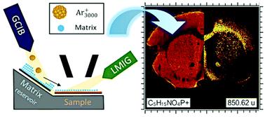
Sensitivity to molecular ions remains a limiting factor for high resolution imaging mass spectrometry of organic and biological materials. Here, we investigate a variant of matrix-enhanced secondary ion mass spectrometry in which the transfer of matrix molecules to the analyte sample is carried out in situ (in situ ME-SIMS). This approach is therefore compatible with both 2D and 3D imaging by SIMS. In this exploratory study, nanoscale matrix layers were sputter-transferred inside our time-of-flight (ToF)-SIMS to a series of thin films of biomolecules (proteins, sugars, lipids) adsorbed on silicon, and the resulting layers were analyzed and depth-profiled. For this purpose, matrix molecules were desorbed from a coated target (obtained by drop-casting or sublimation) using 10 keV Ar3000+ ion beam sputtering, followed by redeposition on a collector carrying the sample to be analyzed. After evaluating the quality of the transfer of six different matrices on bare Si collectors, α-cyano-4-hydroxycinnamic acid (CHCA) was selected for further experiments. The mass spectra and depth profiles obtained from the organic layer prior to and after the sputter-transfer of CHCA were compared, along with those obtained from regular ME-SIMS samples (dried droplets) and, finally, with MALDI data for the same matrix–analyte combinations. Signal amplification factors were calculated by dividing the integrated molecular intensities obtained with or without matrix transfer. While the amplification factors are between 0.5 and 2 for molecules already detected with high intensities in SIMS, such as cholesterol or human angiotensin, other compounds show very large integrated signal amplification, even above two orders of magnitude. This is the case for D-glucose and cardiolipin, for which the molecular ion intensity is low (or very low) under normal SIMS analysis conditions. For such low ionization probability compounds, the beneficial effect of the matrix is unquestionable. Test experiments on mouse brain tissue sections also indicate signal enhancement with the matrix, especially for high mass lipid ions.
中文翻译:

使用原位基质增强二次离子质谱法改进薄膜中的生物分子分析
对分子离子的敏感性仍然是有机和生物材料高分辨率成像质谱的限制因素。在这里,我们研究了基质增强二次离子质谱法的一种变体,其中基质分子向分析物样品的转移是原位进行的(原位ME-SIMS)。因此,这种方法与 SIMS 的 2D 和 3D 成像兼容。在这项探索性研究中,纳米级基质层在我们的飞行时间 (ToF)-SIMS 内被溅射转移到一系列吸附在硅上的生物分子(蛋白质、糖类、脂质)薄膜,并对所得层进行分析和深度剖析。为此,使用 10 keV Ar 从涂层目标(通过滴铸或升华获得)上解吸基质分子3000 +离子束溅射,然后再沉积在携带待分析样品的收集器上。在评估了六种不同基质在裸硅收集器上的转移质量后,选择了 α-氰基-4-羟基肉桂酸 (CHCA) 进行进一步的实验。将 CHCA 溅射转移前后从有机层获得的质谱和深度分布与从常规 ME-SIMS 样品(干液滴)获得的质谱和深度分布进行比较,最后与相同基质的 MALDI 数据进行比较–分析物组合。信号放大系数的计算方法是将使用或不使用基质转移获得的积分分子强度相除。虽然在 SIMS 中已经以高强度检测到的分子(例如胆固醇或人血管紧张素)的放大系数在 0.5 到 2 之间,其他化合物显示出非常大的积分信号放大,甚至超过两个数量级。这是为D-葡萄糖和心磷脂,其分子离子强度在正常 SIMS 分析条件下较低(或非常低)。对于这种低电离概率的化合物,基质的有益效果是毋庸置疑的。在小鼠脑组织切片上的测试实验也表明基质的信号增强,特别是对于高质量脂质离子。
更新日期:2021-09-28
中文翻译:
使用原位基质增强二次离子质谱法改进薄膜中的生物分子分析
对分子离子的敏感性仍然是有机和生物材料高分辨率成像质谱的限制因素。在这里,我们研究了基质增强二次离子质谱法的一种变体,其中基质分子向分析物样品的转移是原位进行的(原位ME-SIMS)。因此,这种方法与 SIMS 的 2D 和 3D 成像兼容。在这项探索性研究中,纳米级基质层在我们的飞行时间 (ToF)-SIMS 内被溅射转移到一系列吸附在硅上的生物分子(蛋白质、糖类、脂质)薄膜,并对所得层进行分析和深度剖析。为此,使用 10 keV Ar 从涂层目标(通过滴铸或升华获得)上解吸基质分子3000 +离子束溅射,然后再沉积在携带待分析样品的收集器上。在评估了六种不同基质在裸硅收集器上的转移质量后,选择了 α-氰基-4-羟基肉桂酸 (CHCA) 进行进一步的实验。将 CHCA 溅射转移前后从有机层获得的质谱和深度分布与从常规 ME-SIMS 样品(干液滴)获得的质谱和深度分布进行比较,最后与相同基质的 MALDI 数据进行比较–分析物组合。信号放大系数的计算方法是将使用或不使用基质转移获得的积分分子强度相除。虽然在 SIMS 中已经以高强度检测到的分子(例如胆固醇或人血管紧张素)的放大系数在 0.5 到 2 之间,其他化合物显示出非常大的积分信号放大,甚至超过两个数量级。这是为D-葡萄糖和心磷脂,其分子离子强度在正常 SIMS 分析条件下较低(或非常低)。对于这种低电离概率的化合物,基质的有益效果是毋庸置疑的。在小鼠脑组织切片上的测试实验也表明基质的信号增强,特别是对于高质量脂质离子。


 京公网安备 11010802027423号