Our official English website, www.x-mol.net, welcomes your feedback! (Note: you will need to create a separate account there.)
Multifunctional superparamagnetic nanoparticles with a fluorescent silica shell for the in vitro study of bio-nano interactions at the subcellular scale
Nanoscale ( IF 6.7 ) Pub Date : 2021-09-27 , DOI: 10.1039/d1nr04582b Lorenzo Cursi 1 , Silvia Vercellino 1, 2 , Mura M McCafferty 1 , Emily Sheridan 1 , Vanya Petseva 1 , Laurent Adumeau 1 , Kenneth A Dawson 1
Nanoscale ( IF 6.7 ) Pub Date : 2021-09-27 , DOI: 10.1039/d1nr04582b Lorenzo Cursi 1 , Silvia Vercellino 1, 2 , Mura M McCafferty 1 , Emily Sheridan 1 , Vanya Petseva 1 , Laurent Adumeau 1 , Kenneth A Dawson 1
Affiliation
|
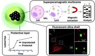
Despite the high level of interest in bio-nano interactions, detailed intracellular mechanisms that govern nanoscale recognition and signalling still need to be unravelled. Magnetic nanoparticles (NPs) are valuable tools for elucidating complex intracellular bio-nano interactions. Using magnetic NPs, it is possible to isolate cell compartments that the particles interact with during intracellular trafficking. Studies at the subcellular scale rely heavily on optical microscopy; therefore, combining the advantages of magnetic recovery with excellent imaging properties to allow intracellular NP tracking is of utmost interest for the nanoscience field. However, it is a challenge to prepare highly magnetic NPs with a suitable fluorescence for the fluorescence imaging techniques typically used for biological studies. Here we present the synthesis of biocompatible multifunctional superparamagnetic multicore NPs with a bright fluorescent silica shell. The incorporation of an organic fluorophore in the silica surrounding the magnetic multicore was optimised to enable the particles to be tracked with the most common imaging techniques. To prevent dye loss resulting from silica dissolution in biological environments, which would reduce the time that the particles could be tracked, we added a thin dense encapsulating silica layer to the NPs which is highly stable in biological media. The synthesised multifunctional nanoparticles were evaluated in cell uptake experiments in which their intracellular location could be clearly identified using fluorescence imaging microscopy, even after 3 days. The magnetic properties of the iron oxide core enabled both efficient recovery of the NPs from the intracellular environment and the extraction of cell compartments involved in their intracellular trafficking. Thus, the NPs reported here provide a promising tool for the study of the processes regulating bio-nano interactions.
中文翻译:

具有荧光二氧化硅壳的多功能超顺磁性纳米粒子,用于亚细胞尺度生物纳米相互作用的体外研究
尽管人们对生物-纳米相互作用非常感兴趣,但仍需要阐明控制纳米级识别和信号传导的详细细胞内机制。磁性纳米粒子 (NPs) 是阐明复杂的细胞内生物纳米相互作用的宝贵工具。使用磁性纳米颗粒,可以隔离颗粒在细胞内运输过程中与之相互作用的细胞隔室。亚细胞尺度的研究严重依赖光学显微镜;因此,将磁恢复的优势与出色的成像特性相结合,允许细胞内 NP 跟踪是纳米科学领域最感兴趣的。然而,为通常用于生物学研究的荧光成像技术制备具有合适荧光的高磁性纳米颗粒是一项挑战。在这里,我们展示了具有明亮荧光二氧化硅壳的生物相容性多功能超顺磁性多核 NP 的合成。在磁性多核周围的二氧化硅中加入有机荧光团经过优化,能够使用最常见的成像技术跟踪颗粒。为了防止生物环境中二氧化硅溶解导致的染料损失,这会减少粒子被跟踪的时间,我们在 NPs 中添加了一层薄而致密的二氧化硅层,该层在生物介质中高度稳定。合成的多功能纳米粒子在细胞摄取实验中进行了评估,其中即使在 3 天后,也可以使用荧光成像显微镜清楚地识别它们的细胞内位置。氧化铁核的磁性既可以有效地从细胞内环境中回收 NP,也可以提取参与其细胞内运输的细胞区室。因此,这里报道的 NPs 为研究调节生物纳米相互作用的过程提供了一个有前途的工具。
更新日期:2021-09-28
中文翻译:
具有荧光二氧化硅壳的多功能超顺磁性纳米粒子,用于亚细胞尺度生物纳米相互作用的体外研究
尽管人们对生物-纳米相互作用非常感兴趣,但仍需要阐明控制纳米级识别和信号传导的详细细胞内机制。磁性纳米粒子 (NPs) 是阐明复杂的细胞内生物纳米相互作用的宝贵工具。使用磁性纳米颗粒,可以隔离颗粒在细胞内运输过程中与之相互作用的细胞隔室。亚细胞尺度的研究严重依赖光学显微镜;因此,将磁恢复的优势与出色的成像特性相结合,允许细胞内 NP 跟踪是纳米科学领域最感兴趣的。然而,为通常用于生物学研究的荧光成像技术制备具有合适荧光的高磁性纳米颗粒是一项挑战。在这里,我们展示了具有明亮荧光二氧化硅壳的生物相容性多功能超顺磁性多核 NP 的合成。在磁性多核周围的二氧化硅中加入有机荧光团经过优化,能够使用最常见的成像技术跟踪颗粒。为了防止生物环境中二氧化硅溶解导致的染料损失,这会减少粒子被跟踪的时间,我们在 NPs 中添加了一层薄而致密的二氧化硅层,该层在生物介质中高度稳定。合成的多功能纳米粒子在细胞摄取实验中进行了评估,其中即使在 3 天后,也可以使用荧光成像显微镜清楚地识别它们的细胞内位置。氧化铁核的磁性既可以有效地从细胞内环境中回收 NP,也可以提取参与其细胞内运输的细胞区室。因此,这里报道的 NPs 为研究调节生物纳米相互作用的过程提供了一个有前途的工具。


 京公网安备 11010802027423号