Our official English website, www.x-mol.net, welcomes your
feedback! (Note: you will need to create a separate account there.)
X-ray excited luminescence spectroscopy and imaging with NaGdF4:Eu and Tb
RSC Advances ( IF 3.9 ) Pub Date : 2021-09-24 , DOI: 10.1039/d1ra05451a Meenakshi Ranasinghe 1 , Md Arifuzzaman 1 , Apeksha C Rajamanthrilage 1 , W R Willoughby 2 , Ashley Dickey 1 , Colin McMillen 1 , Joseph W Kolis 1 , Mark Bolding 2 , Jeffrey N Anker 1
RSC Advances ( IF 3.9 ) Pub Date : 2021-09-24 , DOI: 10.1039/d1ra05451a Meenakshi Ranasinghe 1 , Md Arifuzzaman 1 , Apeksha C Rajamanthrilage 1 , W R Willoughby 2 , Ashley Dickey 1 , Colin McMillen 1 , Joseph W Kolis 1 , Mark Bolding 2 , Jeffrey N Anker 1
Affiliation
|
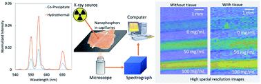
X-ray excited optical luminescence from nanophosphors can be used to selectively generate light in tissue for imaging and stimulating light-responsive materials and cells. Herein, we synthesized X-ray scintillating NaGdF4:Eu and Tb nanophosphors via co-precipitate and hydrothermal methods, encapsulated with silica, functionalized with biotin, and characterized by X-ray excited optical luminescence spectroscopy and imaging. The nanophosphors synthesized by co-precipitate method were ∼90 and ∼106 nm in diameter, respectively, with hydrothermally synthesized particles showing the highest luminescence intensity. More importantly, we investigated the effect of thermal annealing/calcination on the X-ray excited luminescence spectra and intensity. At above 1000 °C, the luminescence intensity increased, but particles fused together. Coating with a 15 nm thick silica shell prevented particle fusion and enabled silane-based chemical functionalization, although luminescence decreased largely due to the increased mass of non-luminescent material. We observed an increase in luminesce intensity with temperature until at 400 °C. At above 600 °C, NaGdF4:Eu@SiO2 converts to NaGd9Si6O26:Eu, an X-ray scintillator brighter than annealed NPs at 400 °C and dimmer than NPs synthesized using the hydrothermal method. The particles generate light through tissue and can be selectively excited using a focused X-ray source for imaging and light generation applications. The particles also act as MRI contrast agents for multi-modal localization.
中文翻译:

X 射线激发发光光谱和 NaGdF4:Eu 和 Tb 成像
纳米磷光体的 X 射线激发光学发光可用于选择性地在组织中产生光,用于成像和刺激光响应材料和细胞。在此,我们通过共沉淀和水热方法合成了X射线闪烁NaGdF 4 :Eu和Tb纳米磷光体,用二氧化硅封装,用生物素功能化,并通过X射线激发光学发光光谱和成像进行表征。共沉淀法合成的纳米荧光粉直径分别为~90 nm和~106 nm,其中水热合成颗粒显示出最高的发光强度。更重要的是,我们研究了热退火/煅烧对 X 射线激发发光光谱和强度的影响。在1000℃以上,发光强度增加,但颗粒融合在一起。 15 nm 厚的二氧化硅壳涂层可防止颗粒融合并实现基于硅烷的化学功能化,尽管由于非发光材料质量的增加而导致发光度大幅下降。我们观察到发光强度随着温度的增加而增加,直到 400 °C。在 600 °C 以上,NaGdF 4 :Eu@SiO 2转化为 NaGd 9 Si 6 O 26 :Eu,这是一种 X 射线闪烁体,比 400 °C 下退火的 NP 更亮,并且比使用水热法合成的 NP 更暗。这些粒子产生穿过组织的光,并且可以使用聚焦 X 射线源选择性地激发,用于成像和光产生应用。这些颗粒还充当多模态定位的 MRI 造影剂。
更新日期:2021-09-24
中文翻译:
X 射线激发发光光谱和 NaGdF4:Eu 和 Tb 成像
纳米磷光体的 X 射线激发光学发光可用于选择性地在组织中产生光,用于成像和刺激光响应材料和细胞。在此,我们通过共沉淀和水热方法合成了X射线闪烁NaGdF 4 :Eu和Tb纳米磷光体,用二氧化硅封装,用生物素功能化,并通过X射线激发光学发光光谱和成像进行表征。共沉淀法合成的纳米荧光粉直径分别为~90 nm和~106 nm,其中水热合成颗粒显示出最高的发光强度。更重要的是,我们研究了热退火/煅烧对 X 射线激发发光光谱和强度的影响。在1000℃以上,发光强度增加,但颗粒融合在一起。 15 nm 厚的二氧化硅壳涂层可防止颗粒融合并实现基于硅烷的化学功能化,尽管由于非发光材料质量的增加而导致发光度大幅下降。我们观察到发光强度随着温度的增加而增加,直到 400 °C。在 600 °C 以上,NaGdF 4 :Eu@SiO 2转化为 NaGd 9 Si 6 O 26 :Eu,这是一种 X 射线闪烁体,比 400 °C 下退火的 NP 更亮,并且比使用水热法合成的 NP 更暗。这些粒子产生穿过组织的光,并且可以使用聚焦 X 射线源选择性地激发,用于成像和光产生应用。这些颗粒还充当多模态定位的 MRI 造影剂。


 京公网安备 11010802027423号