当前位置:
X-MOL 学术
›
Nano Lett.
›
论文详情
Our official English website, www.x-mol.net, welcomes your
feedback! (Note: you will need to create a separate account there.)
Inside the Protein Corona: From Binding Parameters to Unstained Hard and Soft Coronas Visualization
Nano Letters ( IF 9.6 ) Pub Date : 2021-09-23 , DOI: 10.1021/acs.nanolett.1c02416 Flávia E Galdino 1, 2 , Agustin S Picco 3 , Larissa B Capeletti 1 , Jefferson Bettini 4 , Mateus B Cardoso 1, 2
Nano Letters ( IF 9.6 ) Pub Date : 2021-09-23 , DOI: 10.1021/acs.nanolett.1c02416 Flávia E Galdino 1, 2 , Agustin S Picco 3 , Larissa B Capeletti 1 , Jefferson Bettini 4 , Mateus B Cardoso 1, 2
Affiliation
|
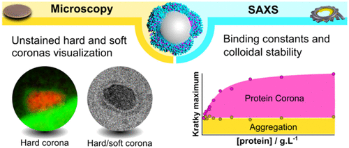
Proteins spontaneously adsorb on nanoparticle surfaces when injected into the bloodstream. It drastically modifies the nanoparticle’s fate and how they interact with organs and cells. Although this protein layer (protein corona) has been widely studied, the robustness of the most employed characterization methods and the visualization of its unstained fractions remain open questions. Here, synchrotron-based small-angle X-ray scattering was used to follow the corona formation and estimate binding parameters. At the same time, transmission electron microscopy under cryogenic conditions associated with cross-correlation image processing and energy-filtered transmission electron microscopy allowed to determine protein corona morphology and thickness together with the visualization of its unstained hard and soft fractions. The above-presented strategy shows tremendous potential for deciphering fundamental protein corona aspects and can contribute to rational medical nanoparticle engineering.
中文翻译:

蛋白质电晕内部:从结合参数到未染色的硬和软电晕可视化
蛋白质在注入血流时会自发吸附在纳米颗粒表面。它极大地改变了纳米颗粒的命运以及它们与器官和细胞的相互作用。尽管该蛋白质层(蛋白质电晕)已被广泛研究,但最常用的表征方法的稳健性及其未染色部分的可视化仍然是悬而未决的问题。在这里,基于同步加速器的小角度 X 射线散射用于跟踪日冕形成并估计结合参数。同时,与互相关图像处理和能量过滤透射电子显微镜相关的低温条件下的透射电子显微镜允许确定蛋白质电晕的形态和厚度,以及其未染色的硬和软部分的可视化。
更新日期:2021-10-13
中文翻译:
蛋白质电晕内部:从结合参数到未染色的硬和软电晕可视化
蛋白质在注入血流时会自发吸附在纳米颗粒表面。它极大地改变了纳米颗粒的命运以及它们与器官和细胞的相互作用。尽管该蛋白质层(蛋白质电晕)已被广泛研究,但最常用的表征方法的稳健性及其未染色部分的可视化仍然是悬而未决的问题。在这里,基于同步加速器的小角度 X 射线散射用于跟踪日冕形成并估计结合参数。同时,与互相关图像处理和能量过滤透射电子显微镜相关的低温条件下的透射电子显微镜允许确定蛋白质电晕的形态和厚度,以及其未染色的硬和软部分的可视化。


 京公网安备 11010802027423号