当前位置:
X-MOL 学术
›
Microsc. Res. Tech.
›
论文详情
Our official English website, www.x-mol.net, welcomes your
feedback! (Note: you will need to create a separate account there.)
Remineralization effects of self-assembling peptide P11-4 associated with three materials on early enamel carious lesions: An in vitro study
Microscopy Research and Technique ( IF 2.0 ) Pub Date : 2021-09-22 , DOI: 10.1002/jemt.23937 Mahtab Memarpour 1 , Faranak Razmjouei 1 , Azade Rafiee 1 , Mehrdad Vossoughi 2
Microscopy Research and Technique ( IF 2.0 ) Pub Date : 2021-09-22 , DOI: 10.1002/jemt.23937 Mahtab Memarpour 1 , Faranak Razmjouei 1 , Azade Rafiee 1 , Mehrdad Vossoughi 2
Affiliation
|
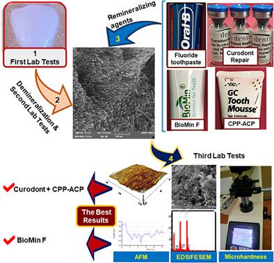
The aim of this study was to assess the remineralization of enamel caries lesions using the self-assembling peptide P11-4 associated with different materials. Artificial early enamel lesions were prepared on 154 primary teeth. The samples were randomly divided into eight groups: (1) control, (2) P11-4, (3) fluoridate toothpaste (FT), (4) P11-4 + FT, (5) casein phosphopeptide–amorphous calcium phosphate (CPP–ACP), (6) P11-4 + CPP–ACP, (7) fluoridate bioactive glass toothpaste (BT), and (8) P11-4 + BT. The surface enamel microhardness (EMH) and energy-dispersive X-ray spectroscopy (EDS) of the teeth were then measured at the baseline, after demineralization, and after 28 days of remineralization. The enamel surfaces were assessed by field emission scanning electron microscopy (FESEM) and atomic force microscopy (AFM). The data were analyzed with one-way analysis of variance (ANOVA) (p < .05). EMH after demineralization was significantly lower than the baseline value (p < .001). The interventions led to an enhanced percentage of EMH recovery (%REMH), which was higher in Groups 6 and 7. There was no significant difference between Groups 3 and 4. Groups 1 and 2 had the lowest %REMH. The mean calcium/phosphate weight percentage ratio of P11-4 was significantly lower than the others (p < .001). The FESEM and AFM images revealed mineral deposition on the eroded enamel and reductions in surface roughness in Groups 5 and 7.
中文翻译:

三种材料相关的自组装肽 P11-4 对早期牙釉质龋损的再矿化作用:一项体外研究
本研究的目的是使用与不同材料相关的自组装肽 P 11 -4 来评估牙釉质龋齿病变的再矿化。在 154 颗乳牙上制备了人工早期牙釉质损伤。将样品随机分为八组:(1)对照,(2)P 11 -4,(3)氟化物牙膏(FT),(4)P 11 -4 + FT,(5)酪蛋白磷酸肽-无定形磷酸钙(CPP–ACP)、(6) P 11 -4 + CPP–ACP、(7) 氟化物生物活性玻璃牙膏 (BT) 和 (8) P 11 -4 + BT。然后在基线、脱矿后和再矿化 28 天后测量牙齿的表面釉质显微硬度 (EMH) 和能量色散 X 射线光谱 (EDS)。通过场发射扫描电子显微镜(FESEM)和原子力显微镜(AFM)评估牙釉质表面。数据采用单因素方差分析 (ANOVA) 进行分析 ( p < .05)。脱矿后的 EMH 显着低于基线值 ( p < .001)。干预措施导致 EMH 恢复百分比 (%REMH) 提高,其中第 6 组和第 7 组较高。第 3 组和第 4 组之间没有显着差异。第 1 组和第 2 组的%REMH 最低。 P 11 -4 的平均钙/磷酸盐重量百分比显着低于其他 ( p < .001)。 FESEM 和 AFM 图像显示第 5 组和第 7 组中被侵蚀的牙釉质上存在矿物质沉积,并且表面粗糙度降低。
更新日期:2021-09-22
中文翻译:
三种材料相关的自组装肽 P11-4 对早期牙釉质龋损的再矿化作用:一项体外研究
本研究的目的是使用与不同材料相关的自组装肽 P 11 -4 来评估牙釉质龋齿病变的再矿化。在 154 颗乳牙上制备了人工早期牙釉质损伤。将样品随机分为八组:(1)对照,(2)P 11 -4,(3)氟化物牙膏(FT),(4)P 11 -4 + FT,(5)酪蛋白磷酸肽-无定形磷酸钙(CPP–ACP)、(6) P 11 -4 + CPP–ACP、(7) 氟化物生物活性玻璃牙膏 (BT) 和 (8) P 11 -4 + BT。然后在基线、脱矿后和再矿化 28 天后测量牙齿的表面釉质显微硬度 (EMH) 和能量色散 X 射线光谱 (EDS)。通过场发射扫描电子显微镜(FESEM)和原子力显微镜(AFM)评估牙釉质表面。数据采用单因素方差分析 (ANOVA) 进行分析 ( p < .05)。脱矿后的 EMH 显着低于基线值 ( p < .001)。干预措施导致 EMH 恢复百分比 (%REMH) 提高,其中第 6 组和第 7 组较高。第 3 组和第 4 组之间没有显着差异。第 1 组和第 2 组的%REMH 最低。 P 11 -4 的平均钙/磷酸盐重量百分比显着低于其他 ( p < .001)。 FESEM 和 AFM 图像显示第 5 组和第 7 组中被侵蚀的牙釉质上存在矿物质沉积,并且表面粗糙度降低。


 京公网安备 11010802027423号